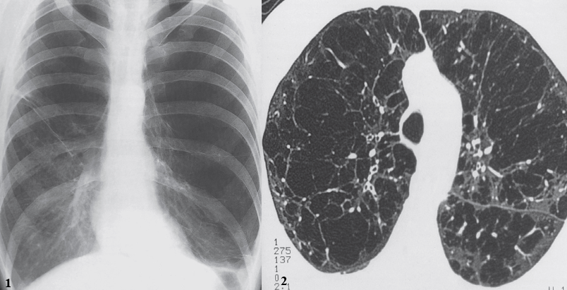
Smoking is the most common cause .
On x-ray chest PA view:
- Increased lung volume.
- The diaphragm is pushed down and becomes low and flat.
- The heart is elongated and narrowed.
- The ribs are widely spaced.
-
The diaphragm is low and flat and the ribs are widely spaced, indicating overinflation of the lungs. - The peripheral vessels in most of the left lung and the upper half of the right lung are small and attenuated, indicating lung destruction.
-
CT showing innumerable bullae.

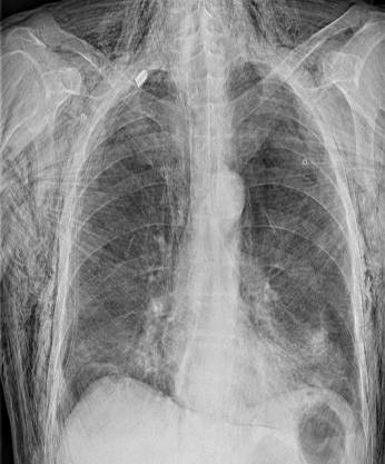
Traumatic/Surgical pneumomediastinum w/ subcutaneous emphysema
X-ray chest (PA view): Extensive subcutaneous emphysema is seen as linear lucencies throughout the soft tissues of the thorax, neck, and upper abdomen. pneumomediastinum is seen along the trachea, main bronchi, and cardiac silhouette .A chest tube is in place, with its tip in the apex of the right hemithorax.
