Role of MRI in ischemic stroke
Cerebral infarction is a kind of stroke that occurs when blood vessels that supply blood in a part of the brain are blocked.
Cerebral infarction actually accounts for 80% of stroke cases worldwide, making it the most common type of stroke.
Sudden onset of focal neurological deficits (e.g., weakness/paralysis, paresthesias, aphasia)
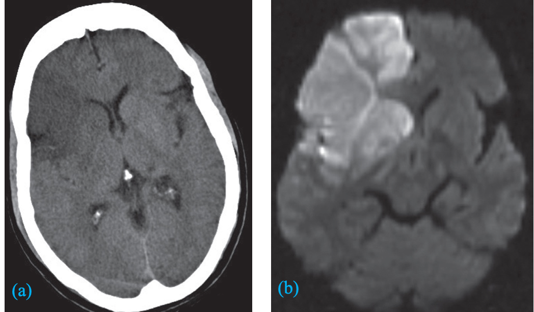
Diffusion-weighted imaging is the most sensitive method for the early detection of an infarct and will show changes within minutes of the onset.
It used in the evaluation of transient ischemic attack and lacunar infarcts.
DWI in stroke.

A) An unenhanced CT image showing low attenuation in the right frontal lobe and basal ganglia .
B) Diffusion-weighted MRI showing bright signal indicating an acute infarct
Noncontrast CT head
All patients with a suspected acute stroke to rule out intracerebral hemorrhage and potential stroke mimics (e.g., tumors) and detect early signs of stroke.
Findings: may be normal or show ischemic changes over time < 2 hours after event: - Usually, no signs of infarction are visible. - In a large artery occlusion, there may be hyperdense occluded vessels (e.g., hyperdense MCA sign).
< 6 hours after the event:
- in some cases, early signs of cytotoxic edema
- Hypodense parenchyma of infarcted region
- Loss of corticomedullary differentiation, e.g. Basal ganglia and insula.
- Effacement of the sulci: e.g., obscuring of the lateral sulcus (also known as Sylvian fissure) due to cortical swelling
12–24 hours after event:
- Hypodense parenchyma starts becoming more clearly demarcated.
***3–5 days after event: ***
- maximum extent of edema and mass effect
2–3 weeks after event:
- (Subacute infarctsƒ) : Infarcted region appears isodense/hypodense.
- Chronic infarcts appear hypodense and well-demarcated, with negative mass effect.
 https://next.amboss.com/us/article/iJ0JFS?q=CT+head+emergency&m=a5XQiA