Pediatrics
Fractures
The most common bones that can be fractured during birth are:
-
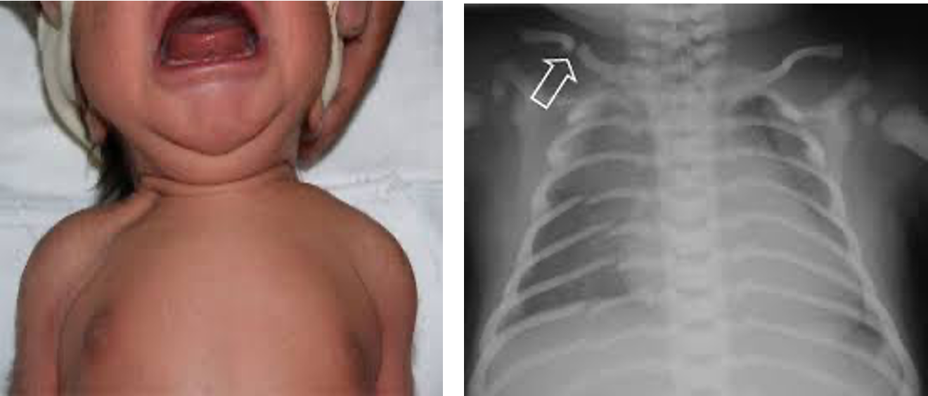
Clavicle:
- The most common fracture.
- A snap may be heard at delivery or the infant may have reduced arm movement on the affected side, or a lump from callus formation later, and unilateral absence of Moro reflex.
- Usually from shoulder dystocia.
- Management: nothing as it heals spontaneously.
-
Skull:
- Linear fractures → observe only.
- Depressed fracture → should be evaluated even if there are no neurological deficits.
-
Long Bones: Humerus/Femur
- Usually, midshaft, occurring at breech deliveries.
- Fracture of the humerus at shoulder dystocia.
- There is a deformity, reduced movement of the limb, and pain on movement.
- They heal rapidly with immobilization.
Radiology
Classification Based on following characteristics: X
- Anatomy; -Location: affected bone (proximal, distal) -Position: diaphysis, metaphysis, epiphysis
- Extent; Complete or incomplete
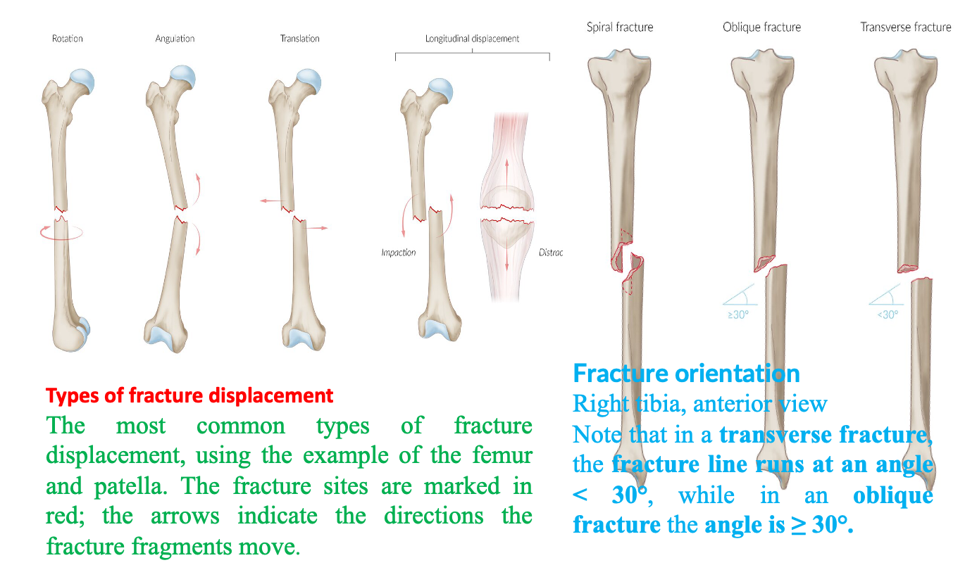
- Orientation; Transverse, Oblique, Spiral
- Displacement;
-Rotated: rotation around the longitudinal axis
-Angulated: angulation of the axis
-Translated: Lateral movement of the bone fragments
-Longitudinal displacement of bone fragments
- Distraction- elongation
- Impaction- shortening
- Fragmentation -Comminuted fracture: more than two fracture lines resulting in multiple bone fragments -Segmental fracture: two fracture lines with a bone fragment between the proximal and distal portions of the bone
- Soft tissue involvement -Closed fracture (simple fracture; does not come into contact with the outside environment) -Open fracture: bone is exposed due to severe soft tissue injury, are associated with a significant risk of infection and poor wound healing.
- Growth plate involvement (pediatric fractures): Salter-Harris fractures
Plain radiography
in skeletal trauma is invaluable in order to:
- Diagnose the presence of a fracture.
- Assess the type of fracture.
- Show displacement and/or angulation of the bone ends before and after treatment.
- Diagnose dislocation or subluxation with or without a fracture.
- Assess healing and complications of fractures.
- Determine whether the fracture has occurred through abnormal bone (pathological fracture).
- Assess soft tissue injury and locate any foreign bodies
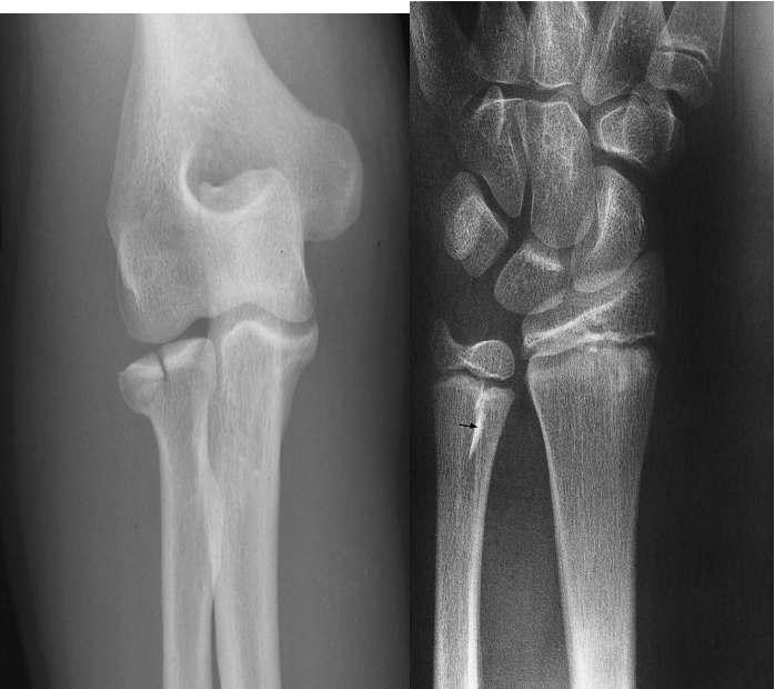 On Plain radiographs fractures may be recognized or suspected by several signs :
A. Fracture of the head of the radius appearing as a lucent line.
B. Fracture of the lower ulnar metaphysis appearing as a sclerotic line (small black arrow).
A fracture is a partial or complete interruption in the continuity of bone. The most common cause is trauma, followed by diseases (e.g., osteoporosis) that result in weakened bone structure.
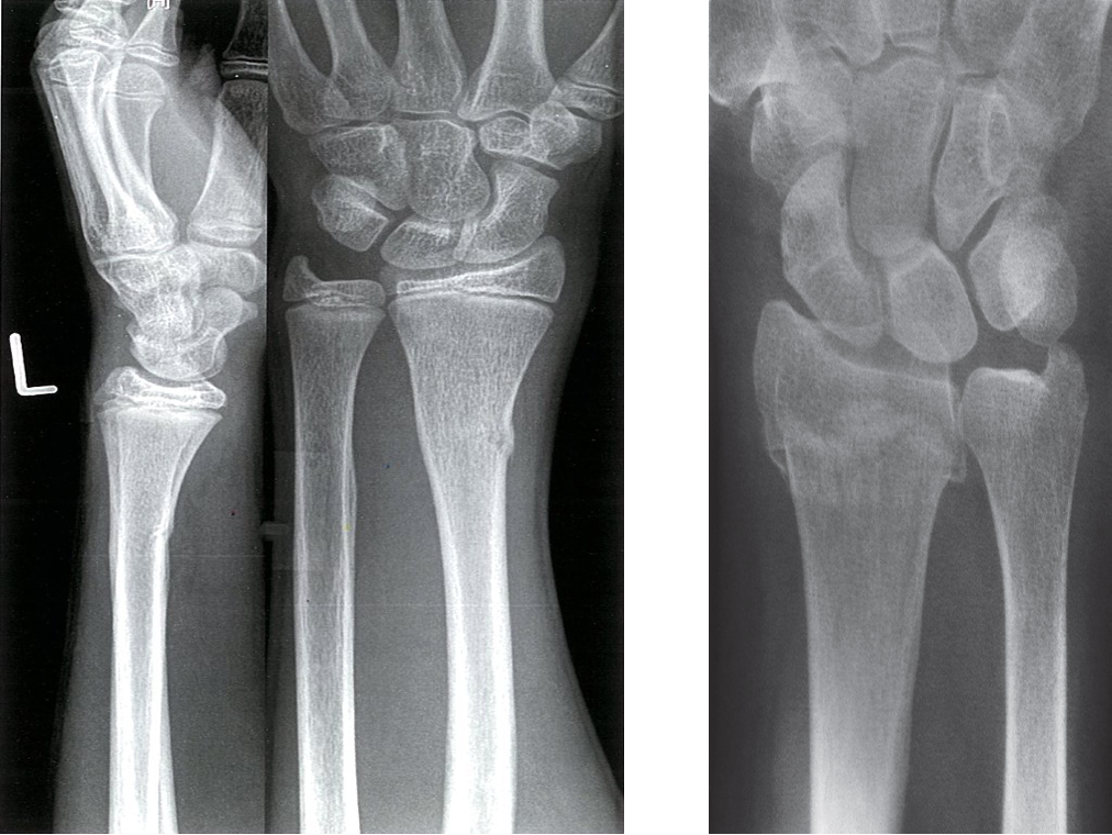 A. X-ray distal left forearm (left: lateral view; right: PA view) of an adolescent - A Greenstick fracture of the distal radius is seen. The cortex on the radial (lateral) side of the fracture is disrupted and buckled . However, the fracture line has not traversed the bone. The ulnar (medial) side of the radius is bowed, but the cortex is intact
B. Step in cortex and interruption of bony trabeculae (arrow) in a Colles’ fracture usually found in elderly
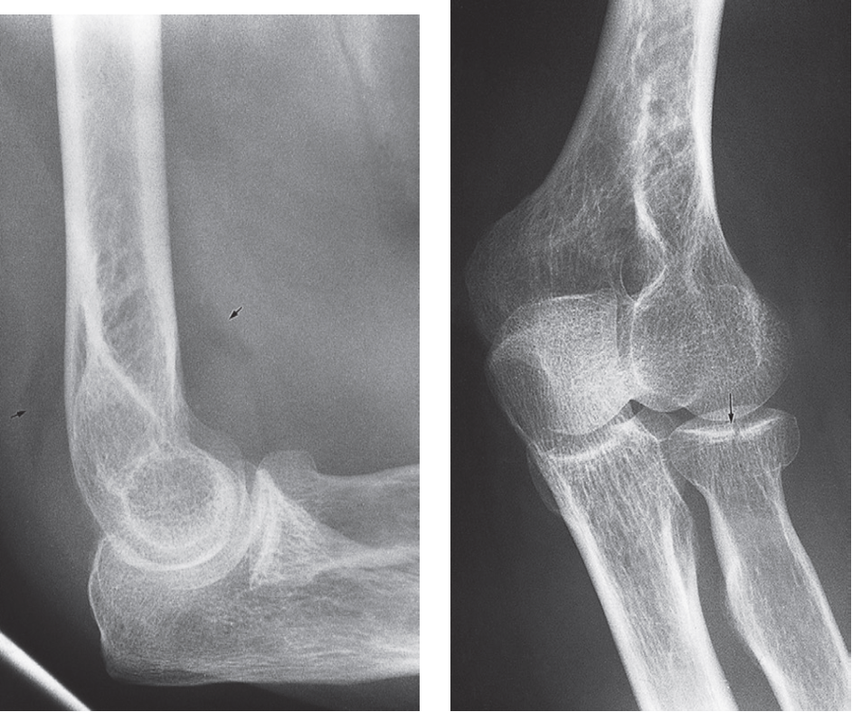 Elbow effusion with fracture of the radial head
Posterior fat pad sign In children suggests a Supracondylar Fractures. In adults, it suggests a radial head fracture.