Obstructing ureteral calculus
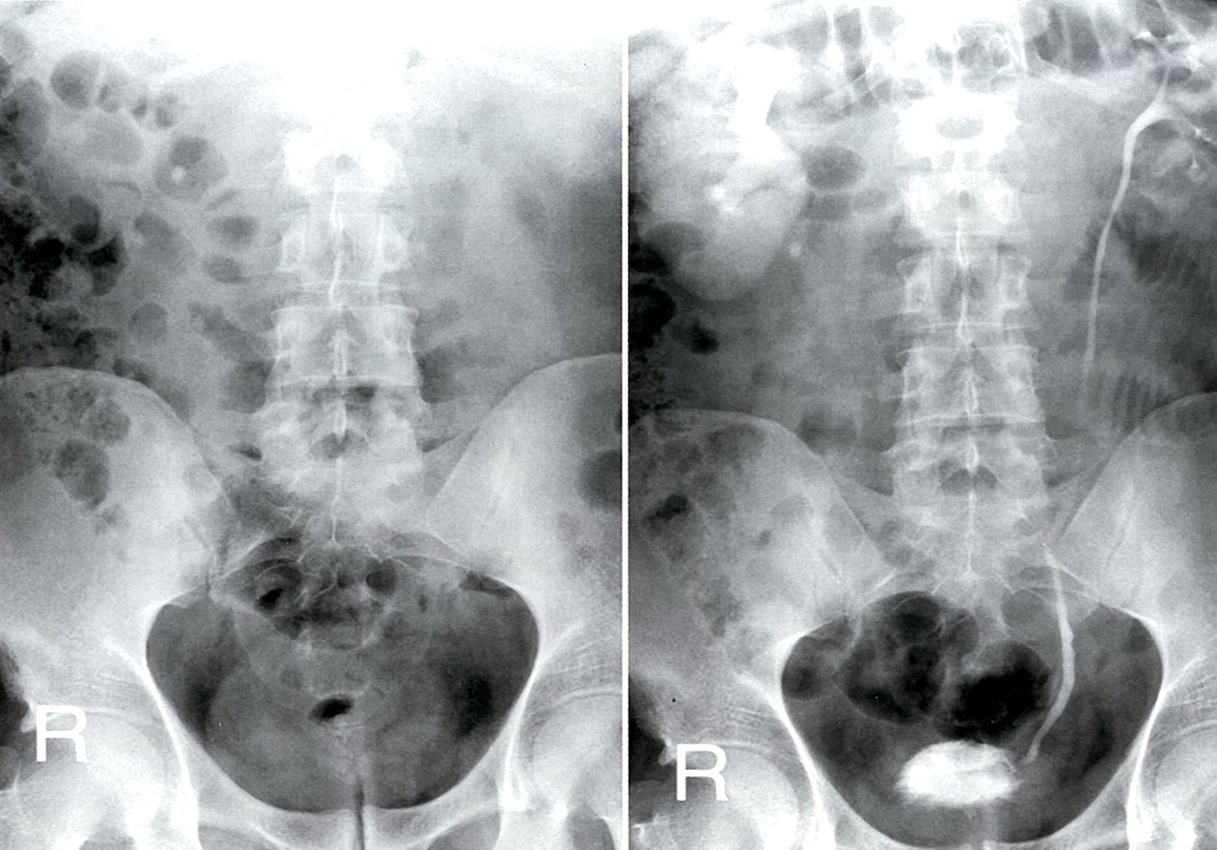
Excretory urography (AP view; left: pre-contrast; right: 2.5 hours post-contrast)
A calcification medial to the lower pole of the right kidney on the pre-contrast radiograph is shown post-contrast to be an obstructing proximal ureteral calculus. The right kidney is enlarged, with persistent enhancement. There is pelvicaliectasis and poor contrast-filling of the right ureter. The left renal collecting system is normal.

Kidney and ureteric stones
Spiral CT without contrast is the investigation of choice for detection of radiolucent stone specially in ureters.
- Calculus size , location, density , and degree of obstruction
- Hydronephrosis and/or hydroureter

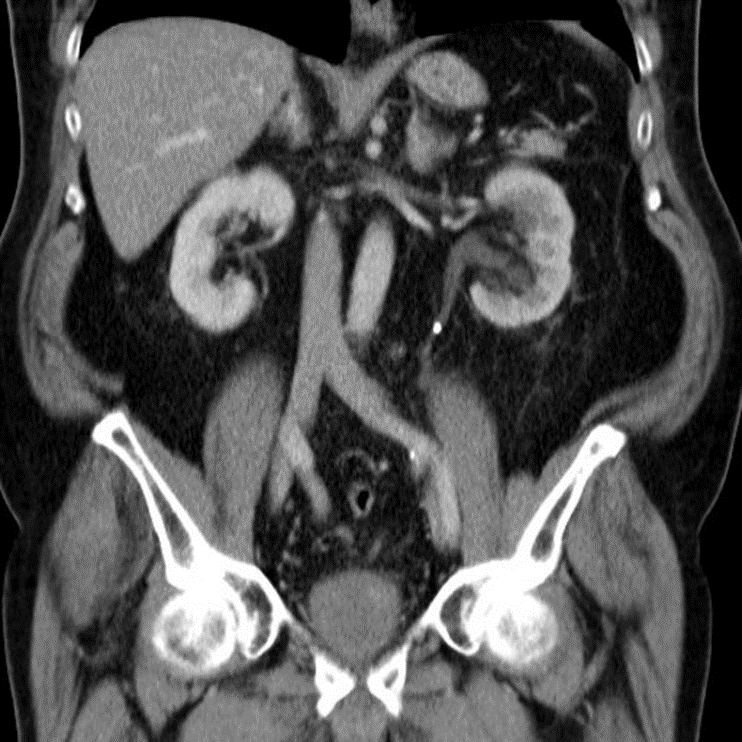
Abdominal CT with IV contrast (venous phase), coronal section
Hyperdense calculi located in the proximal third of the left ureter. As a result, the proximal ureter and the renal pelvis are dilated.

Ultrasound abdomen and pelvis
Indications:
- Suspected nephrolithiasis in patients for whom radiation exposure should be minimized (e.g., pregnant patients, pediatric patients, those with recurrent stones
Findings :
- Obstructive uropathy (e.g., hydronephrosis, hydroureter, perinephric fluid)
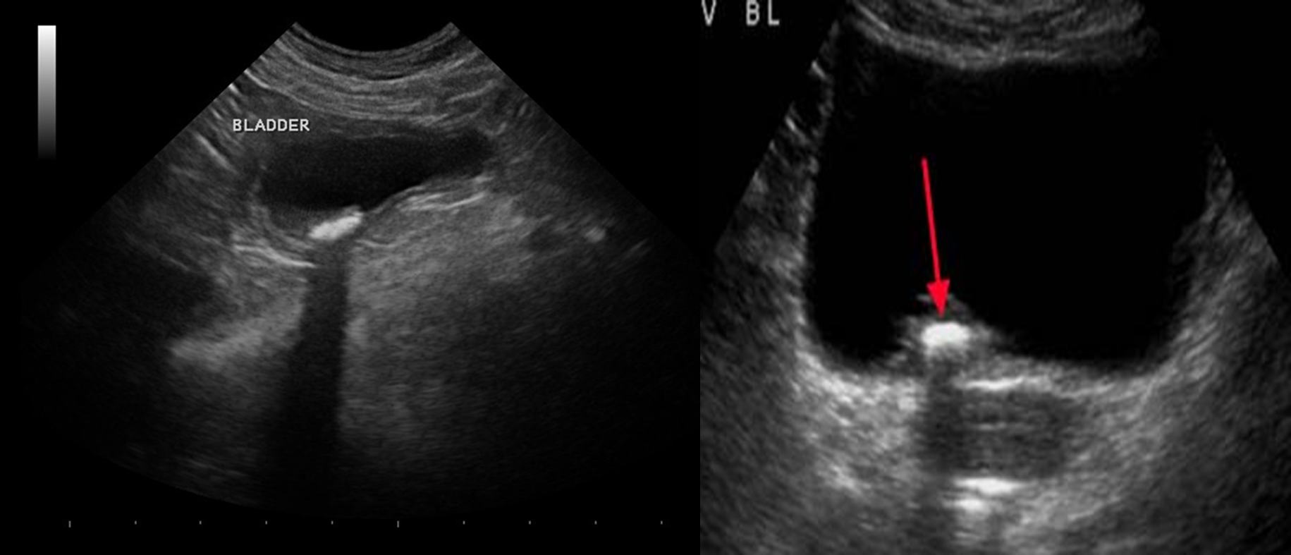
- Stone: hyperechoic with acoustic shadowing
 (1st img) There is a round, hyperechoic lesion at the upper pole of the kidney in the renal parenchyma, which shows a dorsal acoustic shadow and is most likely a kidney stone. The central areas of the kidney show, as far as can be seen in this image, no dilation, which indicates no obstruction.
Bladder calculus
