The Two Layers of the Dermis
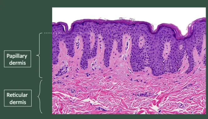
- Papillary Dermis
- Reticular Dermis
Collagen + Ellastine
The Dermis
- The dermis provides a flexible but tough support structure.
- It is between 1-4 mm thick (depending on age and body location), making it much thicker than the epidermis.
- It contains the blood and lymphatic vessels and nerves which supply the skin, as well as sweat glands and hair follicles.
What is the Dermis?
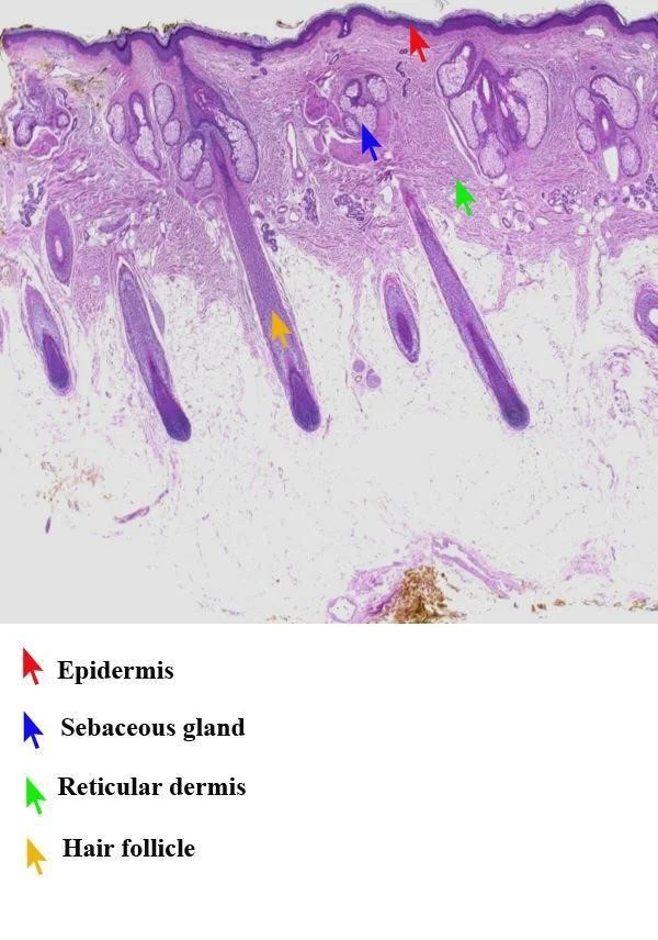
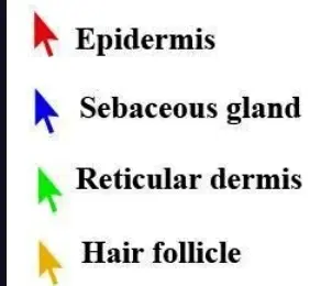
- This is a biopsy from the scalp to show the follicles and sebaceous (oil) glands, found in the dermis.
- There are many hair follicles (yellow arrow) running through the dermis.
- Each follicle has associated sebaceous or oil glands (blue arrow).
- Red arrow – epidermis
- Green arrow – reticular dermis

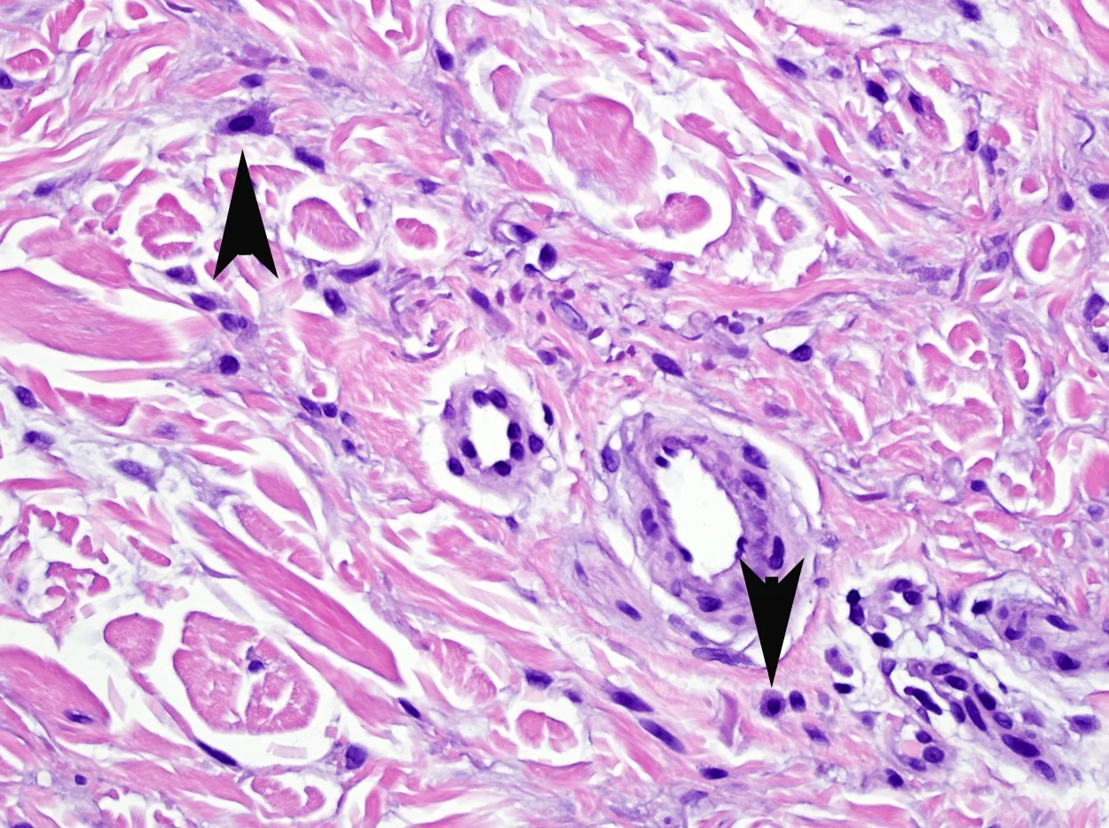
Cells of the Dermis
- Fibroblasts and Mast Cells reside in the dermis
- Fibroblasts are responsible for the synthesis and degradation of connective tissue proteins (elastin).
- They are instrumental in wound healing and scarring.
-
Keloids (abnormal scars) result from uncontrolled synthesis and excessive deposition of collagen at sites of prior dermal injury and wound repair.
-
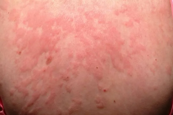
- Mast cells Mast cells are specialized cells that are responsible for immediate-type hypersensitivity reactions in the skin.
- The mast cell is the major effector cell in urticaria, which is a vascular reaction of the skin characterized by wheals surrounded by a red halo or flare.
Outline: Basic Anatomy of the Skin
-
Layers of the Skin
- Epidermis: layers, cell types, and function
- Dermis: layers, cell types, and function
- Subcutis
-
Adnexal Structures
The Subcutis
- The subcutis is the fat layer which separates the dermis from deeper underlying structures such as fascia and muscles.
- The subcutis insulates the body, serves as an energy supply, cushions and protects the skin, and allows for its mobility over underlying structures.
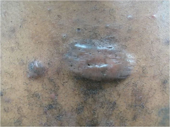
Disorder of the Subcutis
- Erythema nodosum is an example of panniculitis (inflammation of the subcutis).
- Clinically appears as deep-seated erythematous nodules, typically on the shins.
- Erythema nodosum may be idiopathic or a reaction to infections, medication, or an underlying autoimmune disease (e.g., Crohn’s disease).
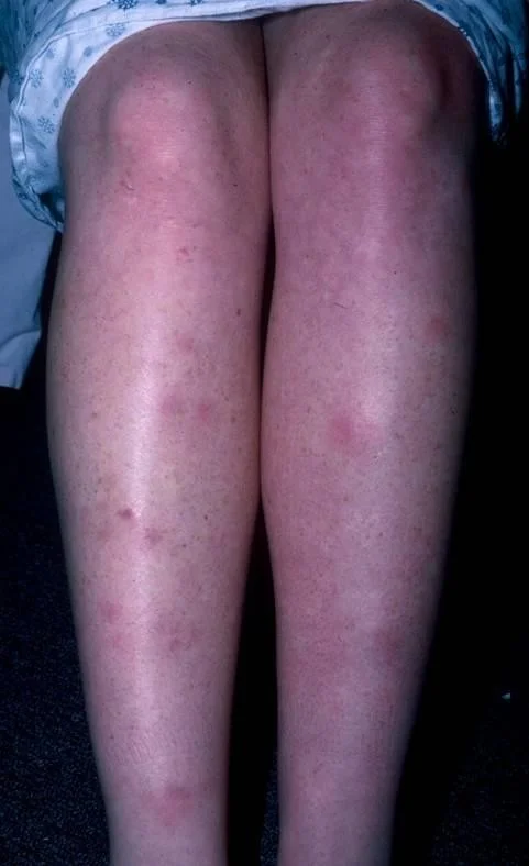 nodules - erythema nodosum
The Pilosebaceous (Hair/Oil Gland) Unit Y
- Adnexal structures include the pilosebaceous unit and eccrine gland.
- Pilosebaceous unit consists:
- A hair follicle
- Sebaceous (oil) glands RR
- Apocrine sweat glands - abnormality happens in soveta reversible acne, pus formation
- An arrector pili muscle (when these contract you get goosebumps)
Apocrine glands are found in the axillary and anogenital areas, which is why we do not see them on this biopsy of the scalp. These glands open directly into the hair follicle.
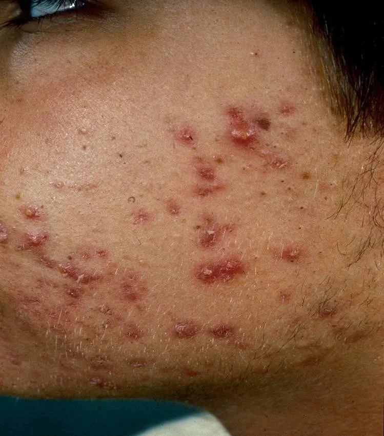
Disorder of Pilosebaceous Unit
- Acne vulgaris is a disorder of the pilosebaceous unit.
Eccrine Glands
- In contrast to apocrine glands, eccrine sweat glands do not involve the hair follicle.
- They open directly onto the skin surface and are present throughout the body.
- Eccrine glands help regulate body temperature by excreting sweat onto the skin surface where cooling evaporation takes place.
- Eccrine glands are sometimes absent, which will predispose a patient to hyperthermia.
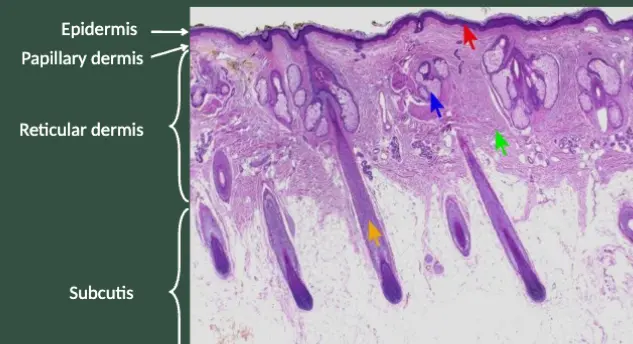
Review: Layers of the Skin
- Epidermis
- Papillary Dermis
- Reticular Dermis
- Subcutis
The epidermis is the purple stripe at the top of the biopsy, and is noted with the red arrow. The reticular dermis is noted with the green arrow. The papillary dermis is the thin bright pink band visible just below the epidermis. The subcutis (fat) is the mostly clear area in the bottom half of the image.
Review Chart: Functions of the Skin Y
| Tissue Layer | Function | Associated Diseases |
|---|---|---|
| Epidermis | Permeability barrier | Atopic dermatitis |
| Epidermis, dermis | Protection from pathogens | Molluscum contagiosum |
| Epidermis, dermis, subcutis | Thermoregulation | Hyperthermia |
| Epidermis | Ultraviolet protection | Albinism |
| Epidermis, dermis, subcutis | Sensation | Diabetic neuropathy |
| Epidermis, dermis | Wound repair/regeneration | Venous stasis ulcer, Keloid |
| Epidermis, dermis, subcutis | Physical appearance | Lipodystrophy |