The most frequently used modality for evaluation of bone and joint disorder
- In bone one view is no view
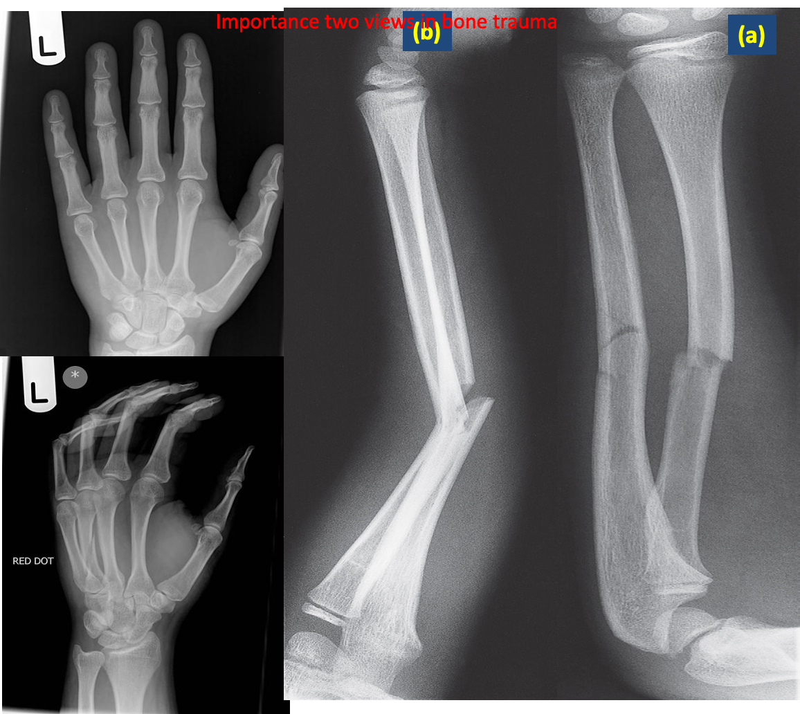
- The radiologist should obtain at least two (2) views of the bone involved at 90° angles to each other .
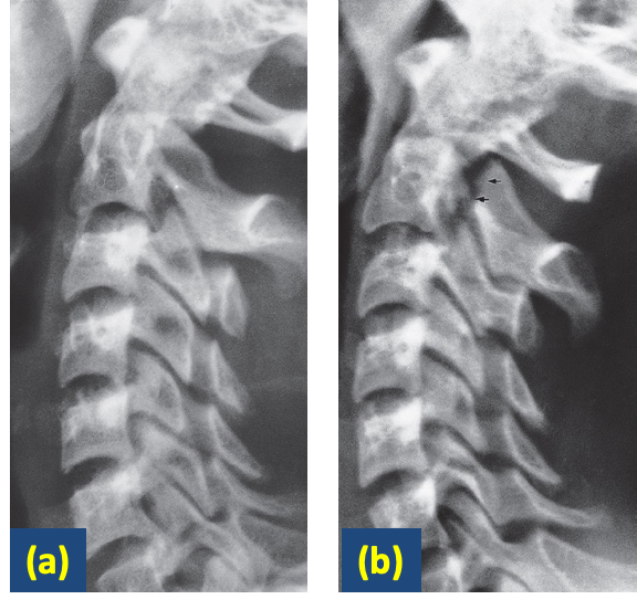
Flexion and extension views. In the cervical spine, injury may cause alteration in the alignment of the posterior borders of the vertebral bodies. This is usually much more obvious on a film taken with the neck flexed
X-ray of the other side. Comparison with the normal side can be useful in the problem case.
Delayed films. This is particularly useful in detecting scaphoid fractures, which may be invisible immediately after the injury.
The signs of bone disease on the radiograph
- decrease in bone density osteopenia. gen
- increase in bone density (sclerosis) gen
- Periosteal reaction
- Alteration in trabecular pattern - osteoporosis & Paget’s Disease
- Alteration in the shape of a bone Acromegaly
- Cortical thickening Chronic Osteomyelitis or healed fracture
Features should be looked for
on plain radiographs & CT when trying to decide the nature of a localized bone lesion:
- Zone of transition. (The edge of any lytic or sclerotic lesion)
- The adjacent cortex malignant tumour or osteomyelitis.
- Expansion enchondroma or fibrous dysplasia.
- Periosteal reaction Codman’s triangle - osteosarcoma
- Calcific densities within the lesion cartilage tumour, neoplasm, infection
Certain lesions tend to occur at certain sites.
-
Osteomyelitis characteristically occurs in the metaphyseal areas, particularly of the knee and lower tibia.
-
giant cell tumors are subarticular in position.
Bone disease diagnosis
When considering the diagnosis and differential diagnosis of bone diseases, it is convenient to divide disorders into five main groups:
- Sclerotic lesions or Solitary lytic.
- Multiple focal lesions, i.e. several discrete lytic or sclerotic lesions or periosteal reactions in one or more bones.
- Generalized lesions where all the bones show diffuse increase or decrease in bone density.
- Those that alter the *trabecular pattern or change its shape
- Fractures
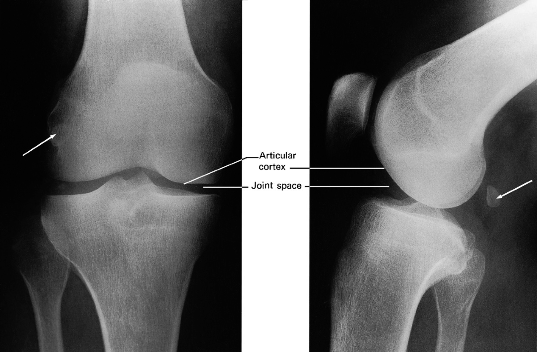
 Normal knee joint. Note the fabella (arrow), a sesamoid bone in the gastrocnemius. The ‘joint space’ consists of articular cartilage and synovial fluid.
 Value of two views for demonstrating the position of fractures.
(a) The fractures of the radius and ulna show little displacement on the frontal projection.
(b) The lateral view, however, shows a marked angulation.
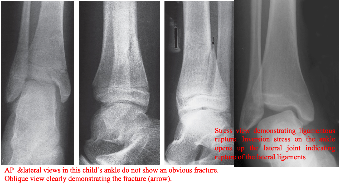
 Injuries may sometimes be invisible even with two views taken at right angles to each other.
Further plain film views
- Different projections, e.g. oblique views
- Stress films: A film taken with a joint under stress; Stress films are helpful in ankle injuries when forced inversion and eversion may show movement of the talus.
 Flexion and extension views demonstrating a fracture.
A) An extension view of the cervical spine does not reveal a fracture.
B) flexion view clearly shows the fracture of the arch of C2.