X

 Basal ganglia hemorrhage
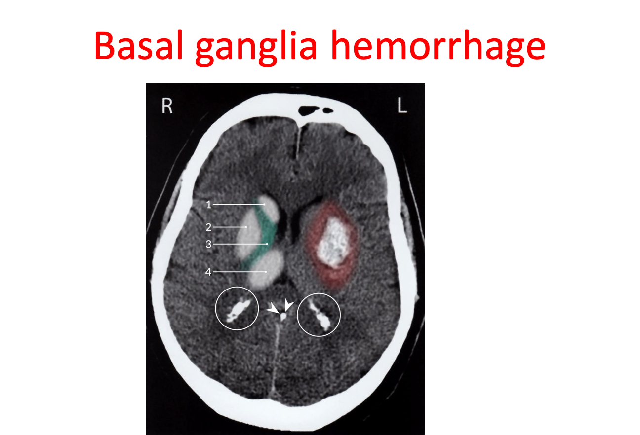
CT head (without contrast; axial plane) of a patient with a history of chronic hypertension
Hyperdense hemorrhage in the left basal ganglia is surrounded by hypodense perifocal edema (red overlay).
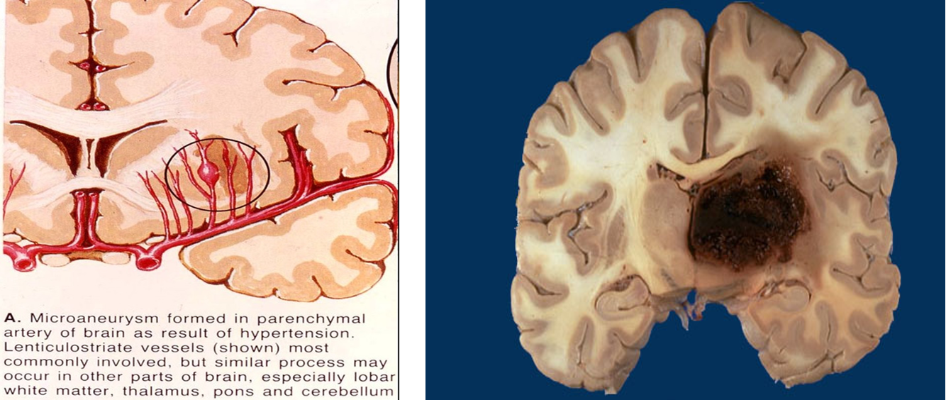
The location is typical for hemorrhage from the lenticulostriate arteries as a result of long-standing hypertension.
1: head of caudate nucleus; 2: putamen; 3 and green overlay: internal capsule; 4: thalamus
Arrowheads: calcified pineal gland; white circles: calcified choroid plexus