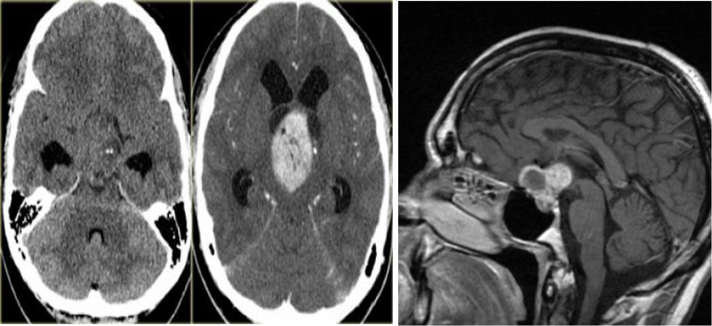
The tumor arises in the suprasellar region and can extend into the intrasellar region.
Bimodal distribution:
- 5–14 years; second peak at 50–75 years
- Most common childhood supratentorial tumor
Imaging: Suprasellar calcified cyst with a lobulated contour

A,B) Craniopharyngioma pre and post contrast
C) MRI head (T1 weighted; with contrast; sagittal of a patient with acute visual loss in the right eye - There is an intrasellar and suprasellar mass with both cystic and solid portions. The solid portions show strong contrast enhancement.