-
Post menopausal endometrium
-
Obstetrics trimesters
-
Fetal wellbeing
-
Detection of intrauterine contraceptive devices
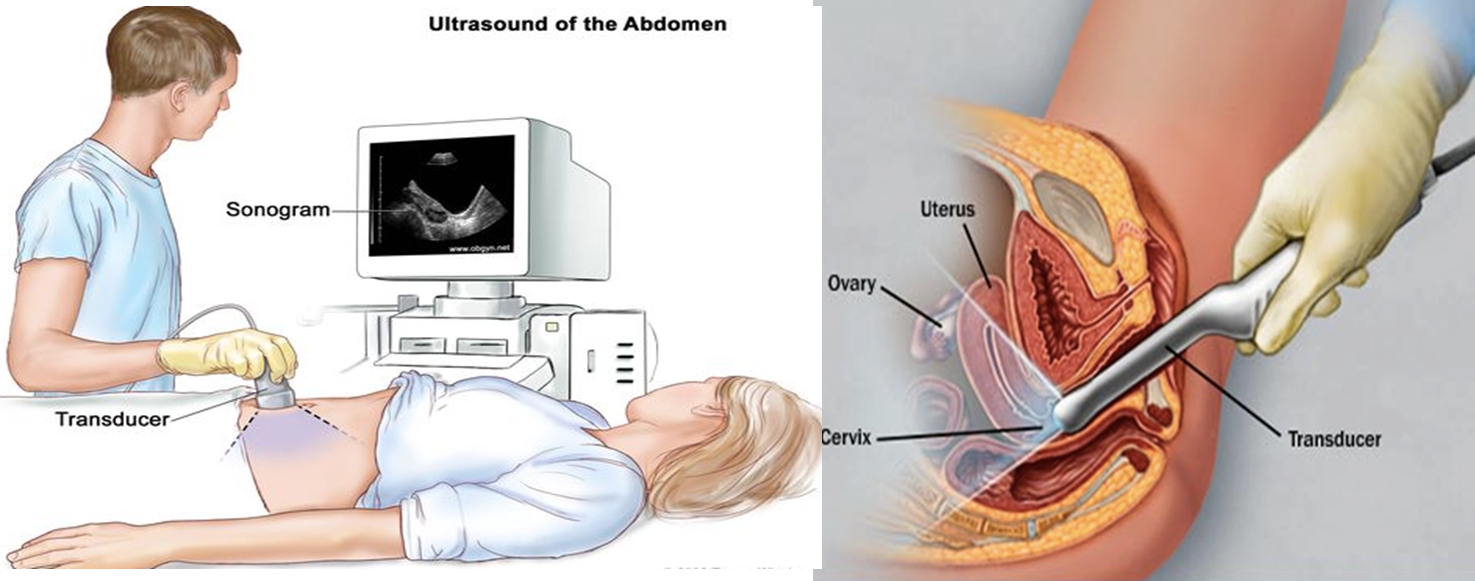
Trans abdominal ultrasound(Need full bladder) vs Trans vaginal ultrasound (Need empty bladder)
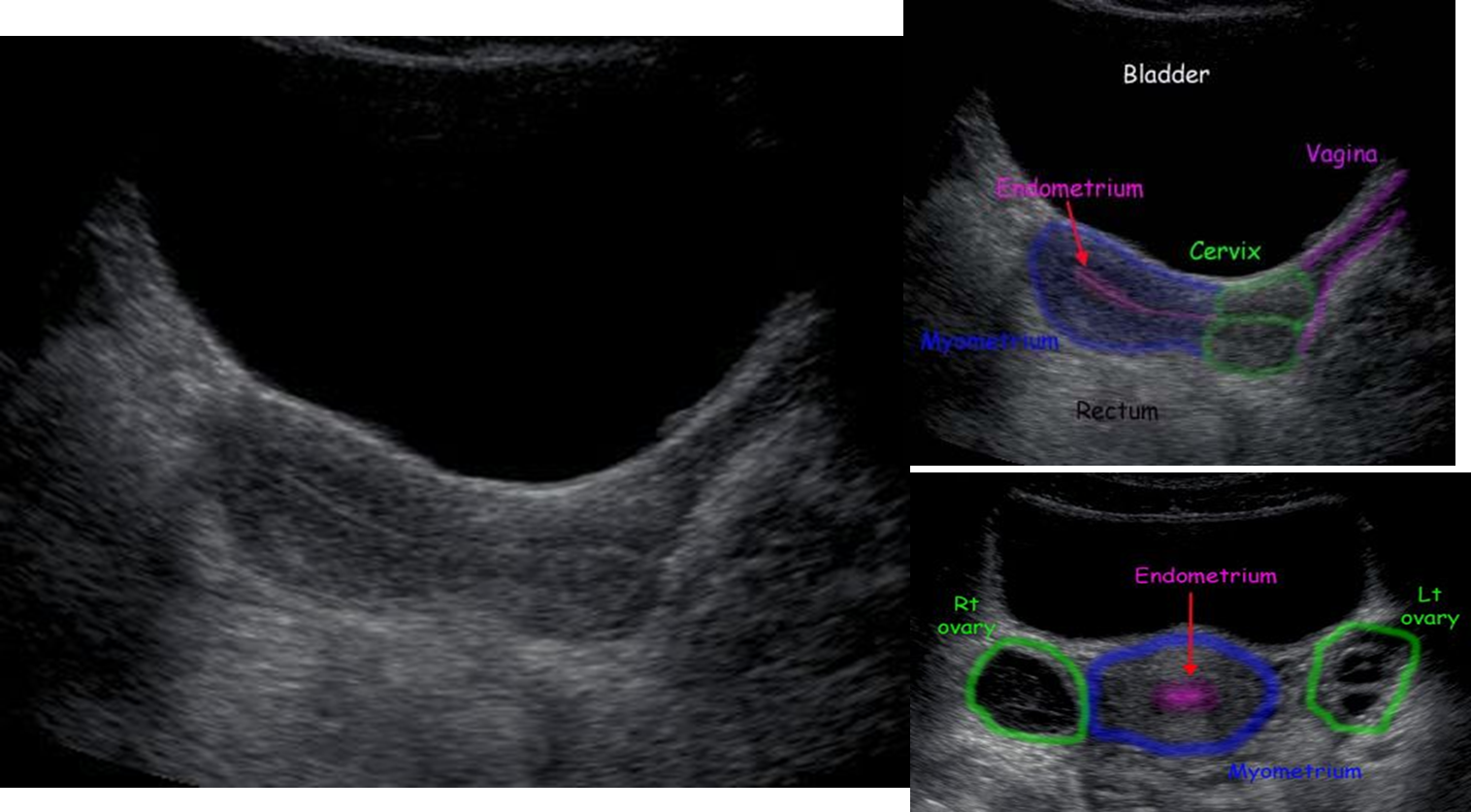
 Normal uterus on trans-abdominal ultrasound Longitudinal and transverse views
- The myometrium shows low level echoes, whereas the endometrial cavity gives a an echogenic linear stripe

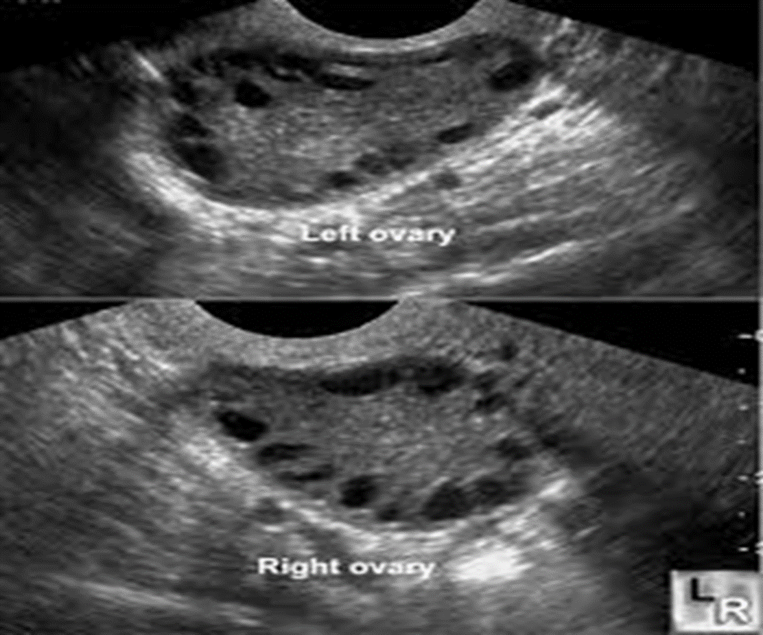
Normal ovary - Ultrasound

- Central echogenic stroma with peripheral anechoic follicles ( 3- 4mm)
- Surrounding hyperechoic Tunica
Placental imaging
-
The placenta is easily evaluated sonographically as well as on MRI.
-
By the ninth week, it is seen as a well-defined intrauterine structure lining the inner wall of a portion of the uterine cavity.
-
Abnormalities related to the placenta may be diagnosed on ultrasound, including placental haemorrhage and placenta previa (placenta remains positioned over the lower uterine segment after the 36th week of pregnancy).
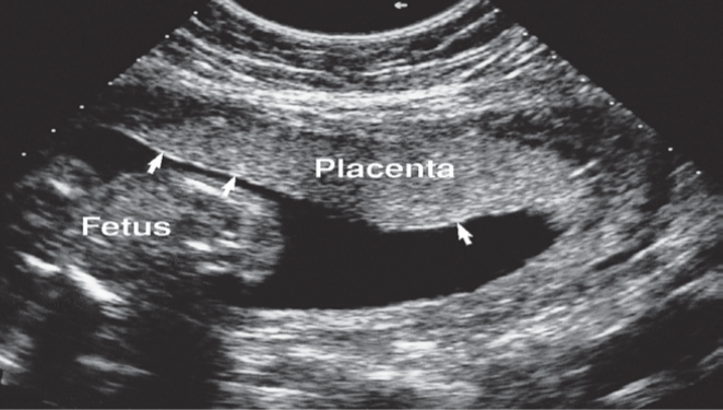
 Placenta seen on a longitudinal scan at 18 weeks’ gestation. The chorionic plate is seen as a thin line of bright echoes (arrows).