Ultrasound is the first line investigation in most patients, providing anatomical information without requiring ionizing radiation or the use of intravenous contrast medium.
-
Evaluate renal parenchyma, bladder, ureters and prostate.
-
Can differentiate between solid and cystic mass, identify simple cyst and hydronephrosis.
-
shows all type of stone
-
Evaluate congenital anomalies.

Normal renal ultrasound Z
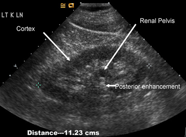
The kidneys should be smooth in outline. The parenchyma surrounds a central echogenic region, known as the central echo complex (the renal sinus), consisting of the pelvicaliceal system, together with the surrounding fat and renal blood vessels.
The normal pelvicaliceal system is not visible within the renal sinus.
The renal cortex generates homogeneous echoes that are of equal or less reflective than those of the adjacent liver or spleen.
-
The renal pyramids are seen as triangular hypoechoic areas adjacent to the renal sinus.
-
The normal adult renal length, measured by ultrasound is 9–12 cm. Renal length varies with age.
-
Normal ureters are not usually visualized due to overlying bowel gas.
-
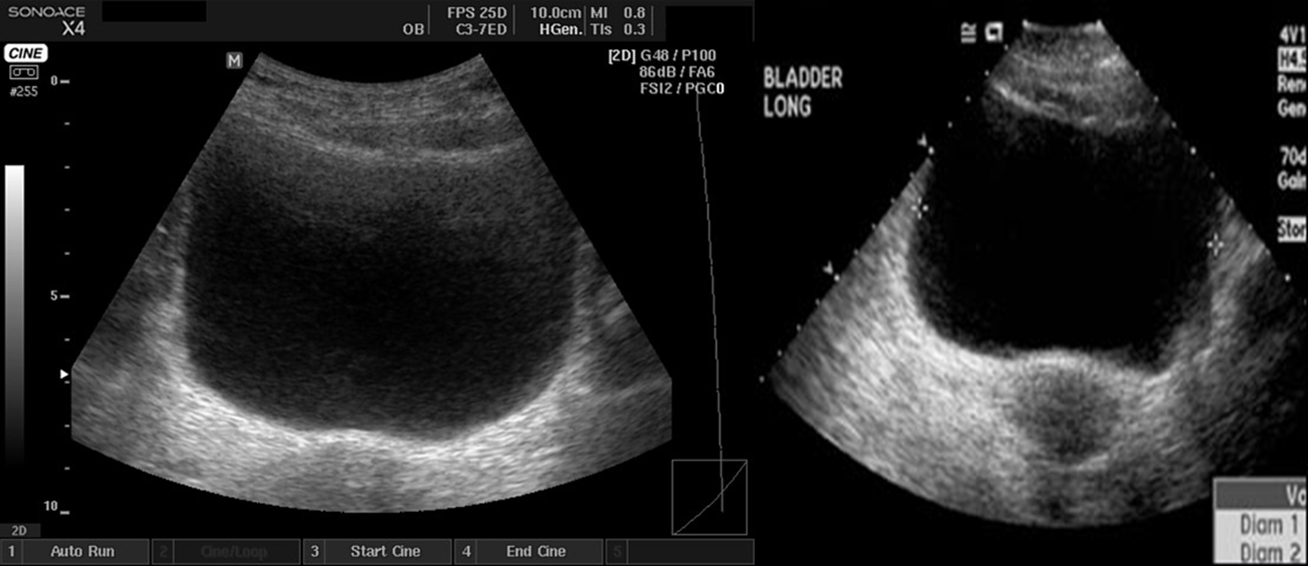
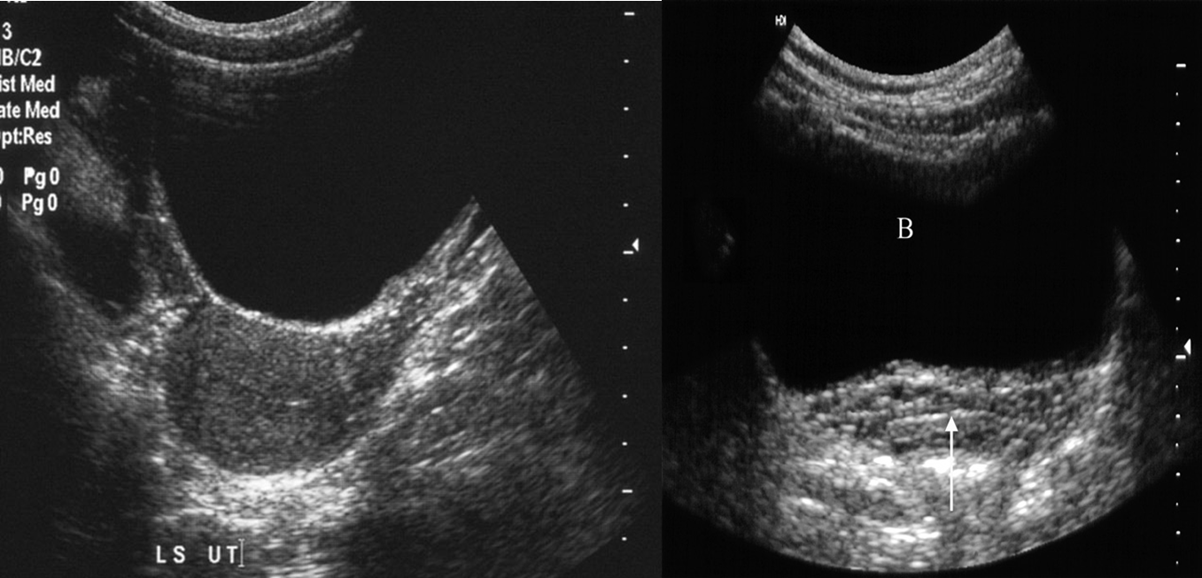
The urinary bladder should be examined in the distended state: the walls should be sharply defined and barely perceptible.

Male

Female
Normal ultrasound of the full bladder (B). Note the smooth thin bladder wall. The vagina lies posteriorly (arrow).
