Brain Imaging
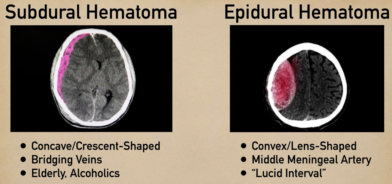
Epidural
- ipsilateral pupil diltation
Treatment: ligation of artery
Subdural
Bridging vein from brain to dural space - commonly elderly/pediatrics
s/s
- chronic
- change in personality
- hx of minor truama
Treatment Purr hole
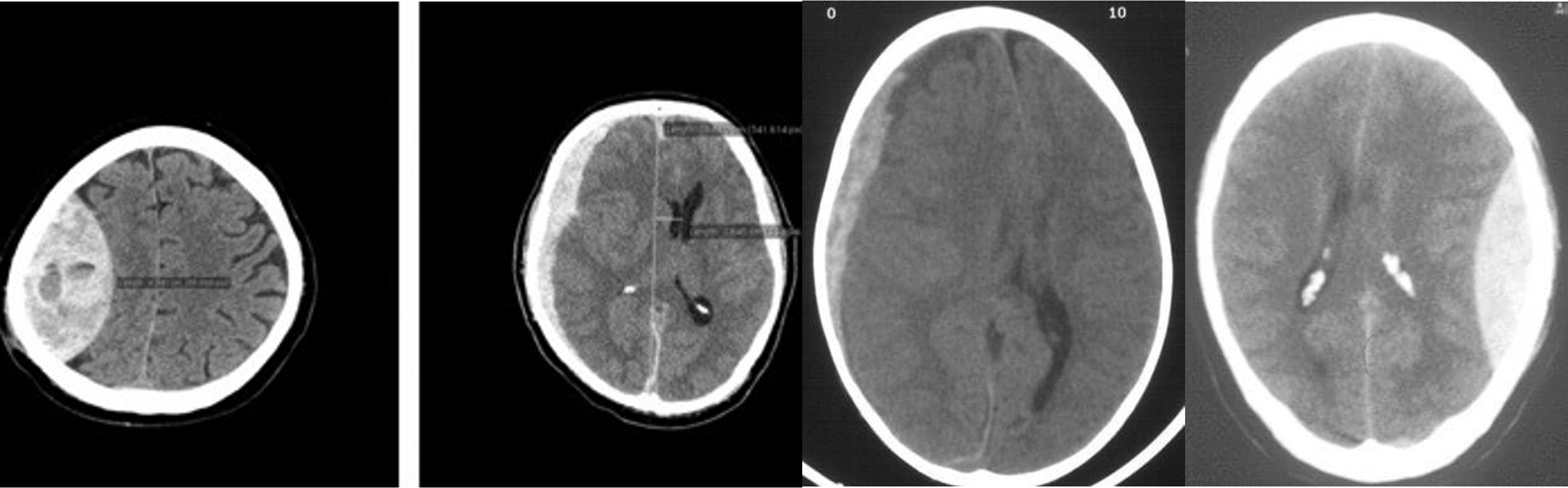
- Epidural - shifted lenticular
- Subdural - crescent
- Subdural -
- Epidural
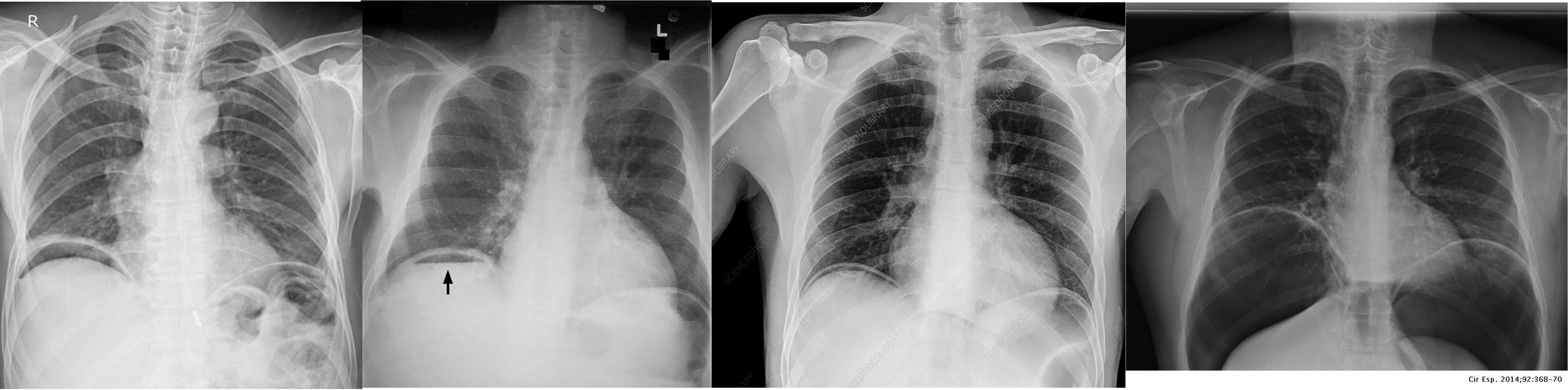
Chest Imaging
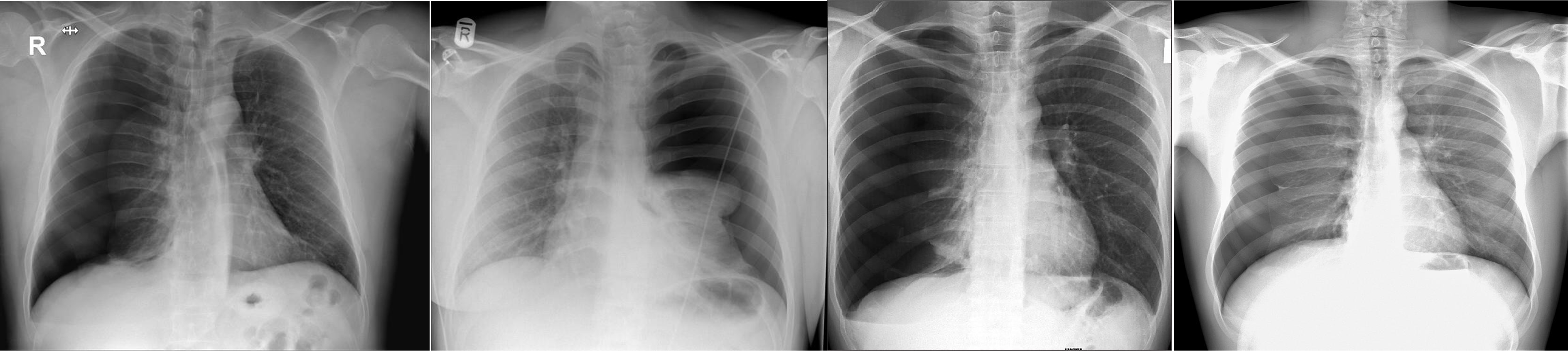
- Pneumothorax - Right lung
- Pneumothorax - Left lung - mediastinum pushed
- Pneumothorax - RT
- Pneumothorax - RT
Abdomen Imaging
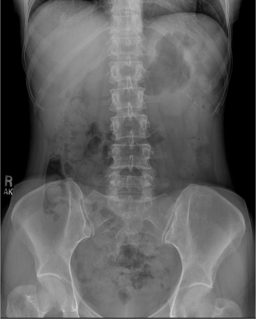 normal abdominal x-ray - normal gas pattern - lower pole kidney - gastric bubble is found - comment on bone
 Free air under diaphragm - most commonly due duodenal perforation - (diff; perforated viscus, peptic ulcer, duodenal, penetrating truama, post surgical 8 days likely to disappear)
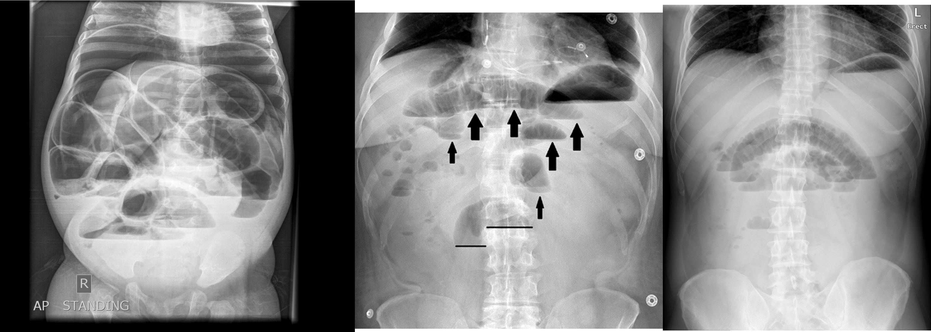
- free air
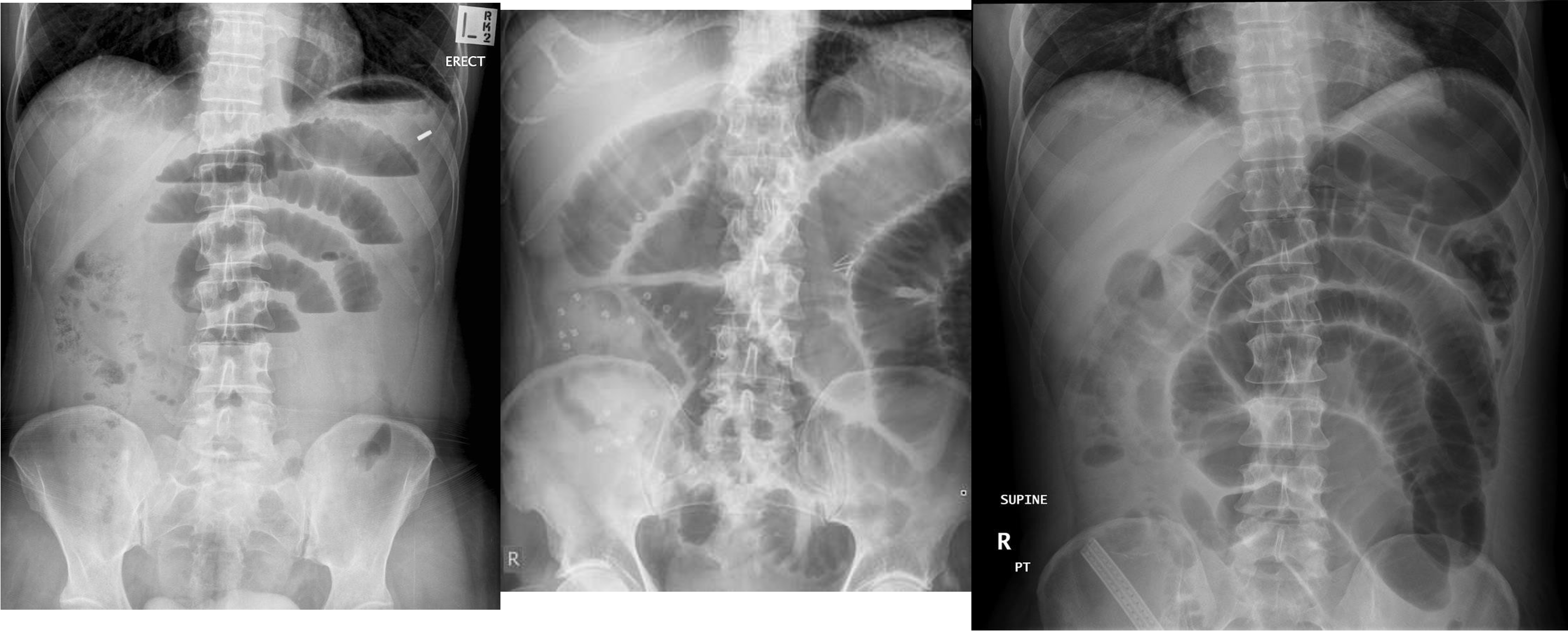
- erect - air fluid levels - should be not more than three - >5 significant 11 found -
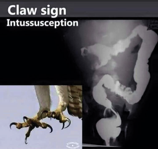
- no free air under diaphgram - 8 fluid level, (evidence of bowel obstruction) (adhesion, hernia, IBD; Crohn, cancer, inturcucception, gallstones)
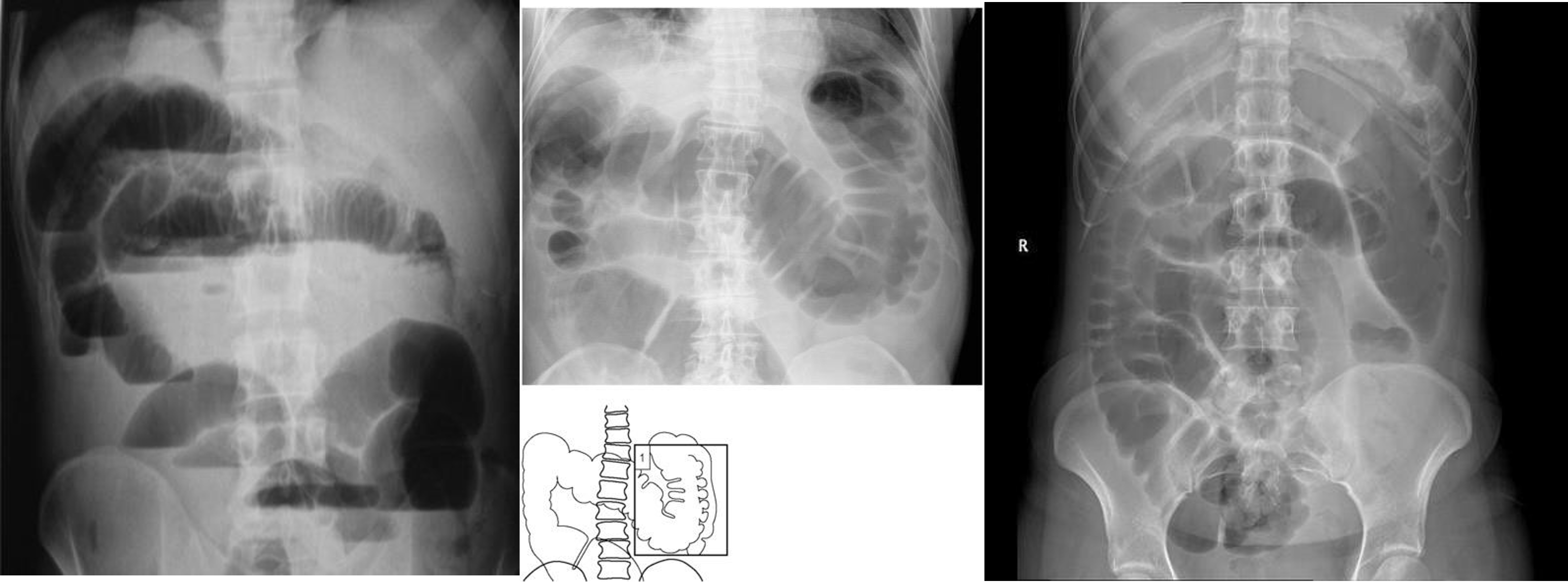
-
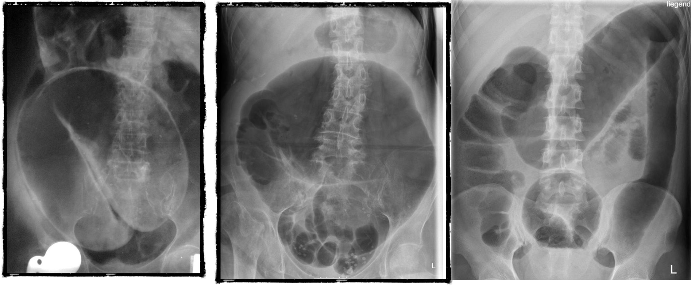
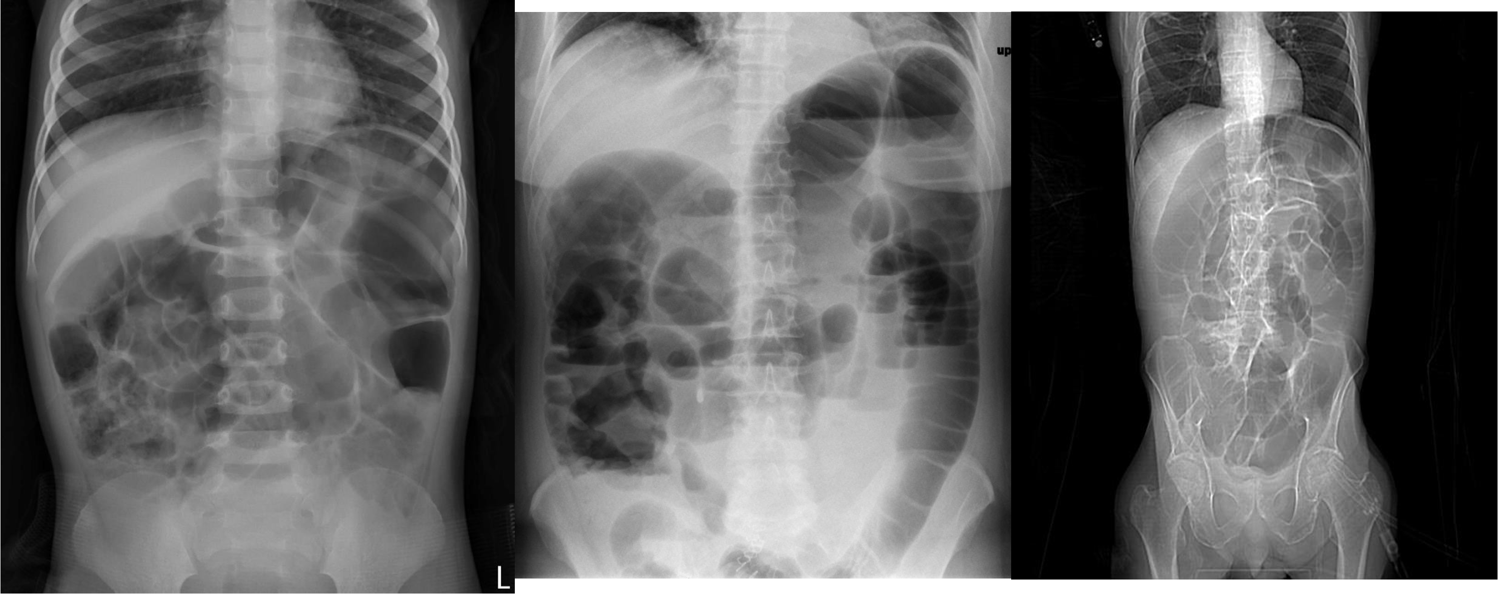
9 air fluids in large bowel - mucosal folds are not completed - highly suggestive of bowel obstruction
- dilated large bowel - mucosal folds are not complete - (diff; cancer, divertuclitis, volvolus, toxic megacolon?)
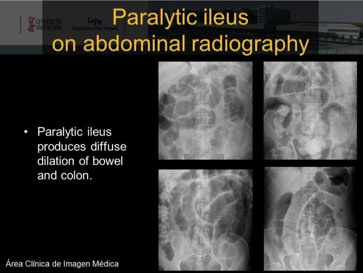
- if both small and large most likely nonmechanical paralytical illeus
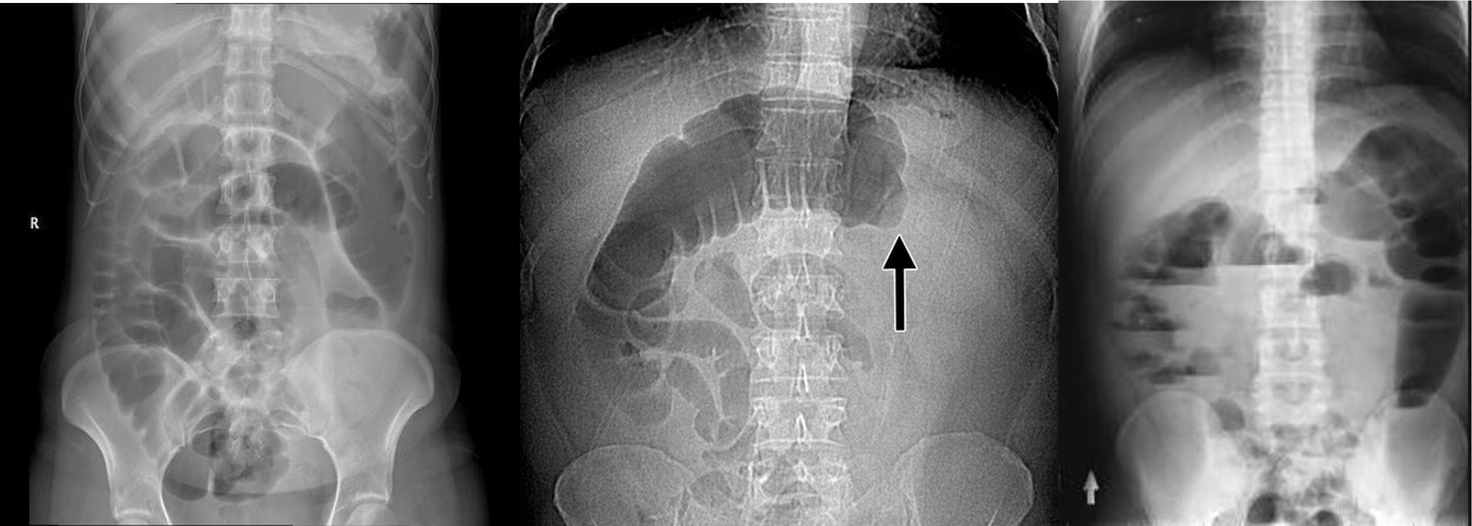 2) Dilated bowel (diff; cancer colon) -
closed loop obstruction from both sides result large bowel most dangerous left side most common cancers
- coffe bean - omega sign - volvulus twisting around its axis - it is closed loop obstruction very dangerous - (treat clonoscopy decompression)
- volvulus
- abdominal x-ray - Dilated colon (diff - cancer colon, ulcerative colittis)
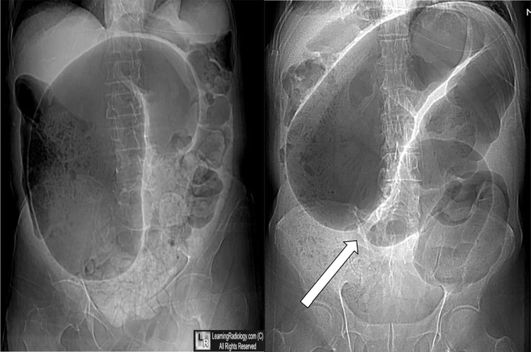
- volvulus
- volvulus
Pyelo
- & 2 Appendicitis - appendicolith/feacolith in young, MALT GALT
- IVP, normal
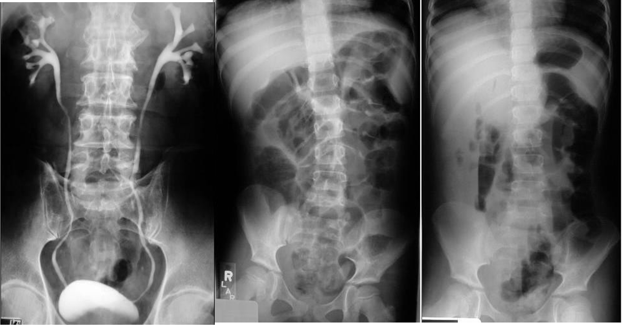
- IVP, Hydrouterer - sharp blunted
- /////
- ////
- 8 air fluid - small intestinal central - intistinal obstruction due adhesion - stack of coins. ++ NG tube aspiration relieve from vomitting
- dilated small bowel obstruction - stack of coins
- small bowel
 1)
2) large bowel - mucosal issnt complete periphery
3)
1)
2) large bowel - mucosal issnt complete periphery
3)
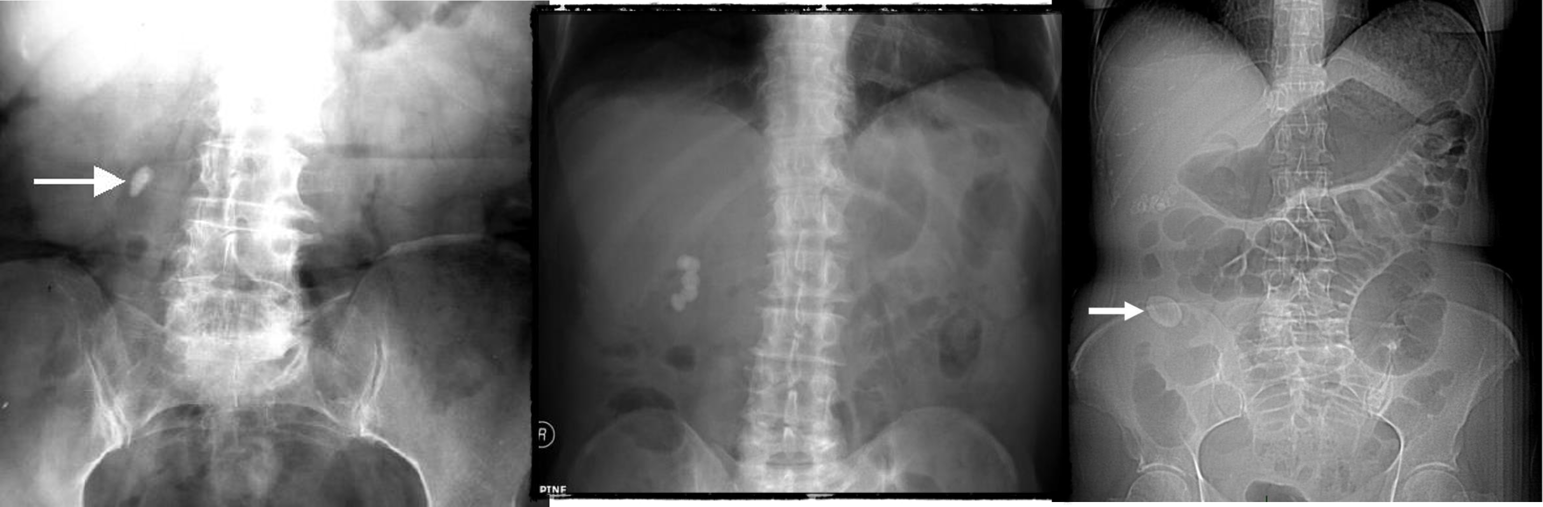
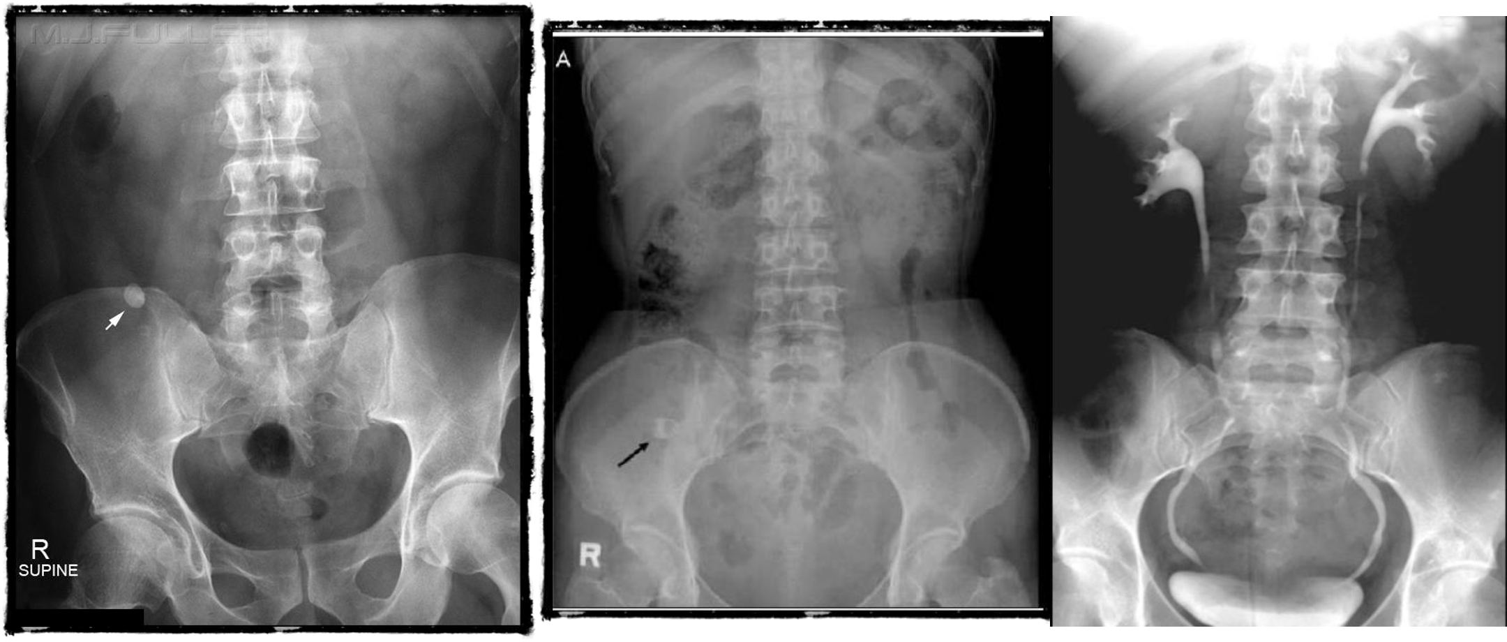
- Uretic stone
- radioopaque shadow on RUQ - renal stone
- Gallstone illeus - radioopaque shadow (regulars triad??)
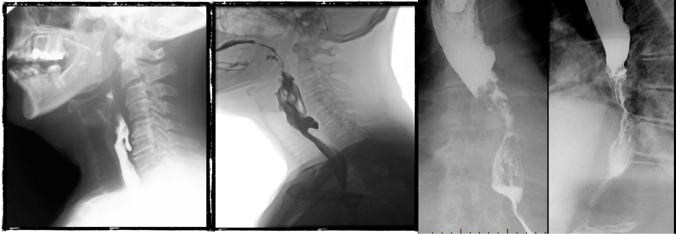
- barium swallow - zenkers diverticulum
- negative
- b swallow - diltation upper, stricture (diff cancer)
- normal (mucosal fold roge)
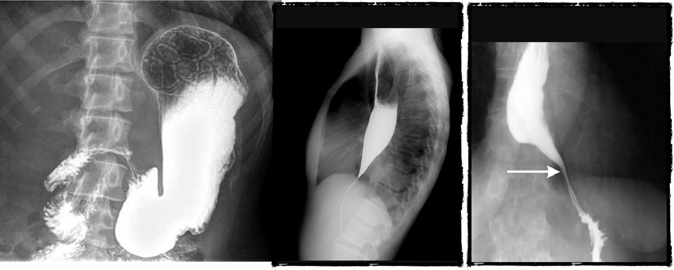
- dilated esophagus, stricture, bear beak/rat tail sign = aclasia
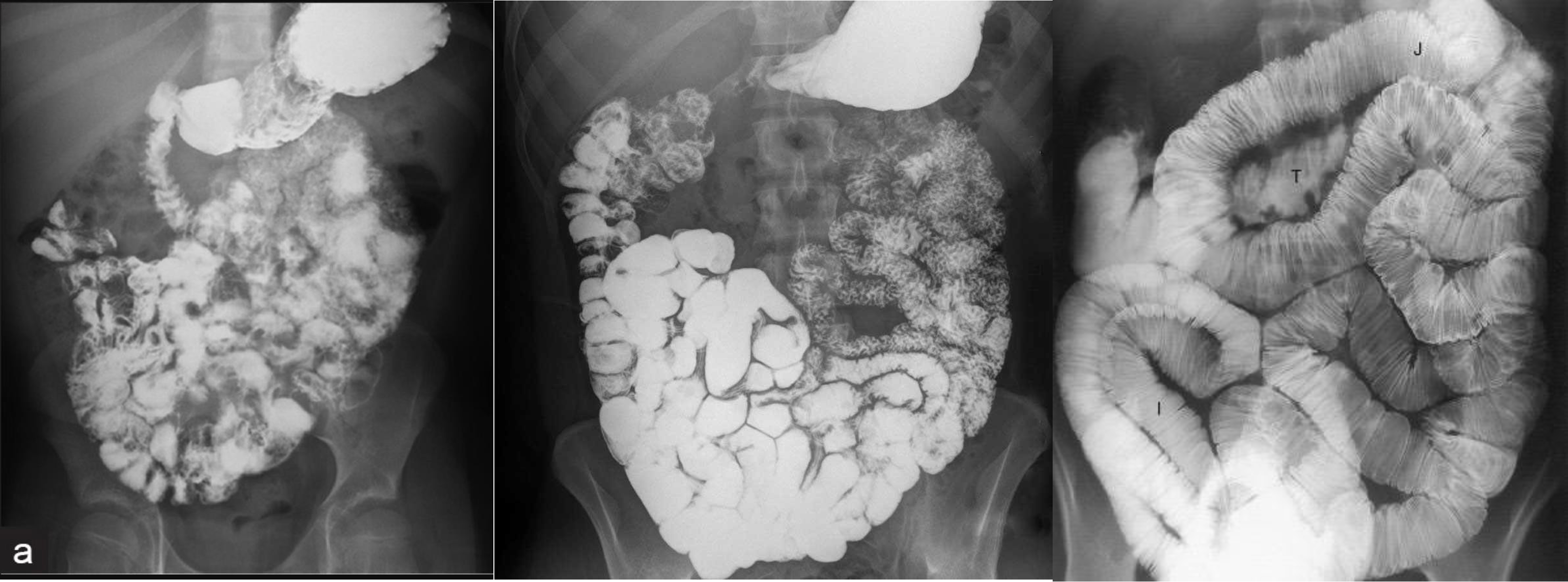
- follow through normal
- follow through (diff celiac; villus atrophy)
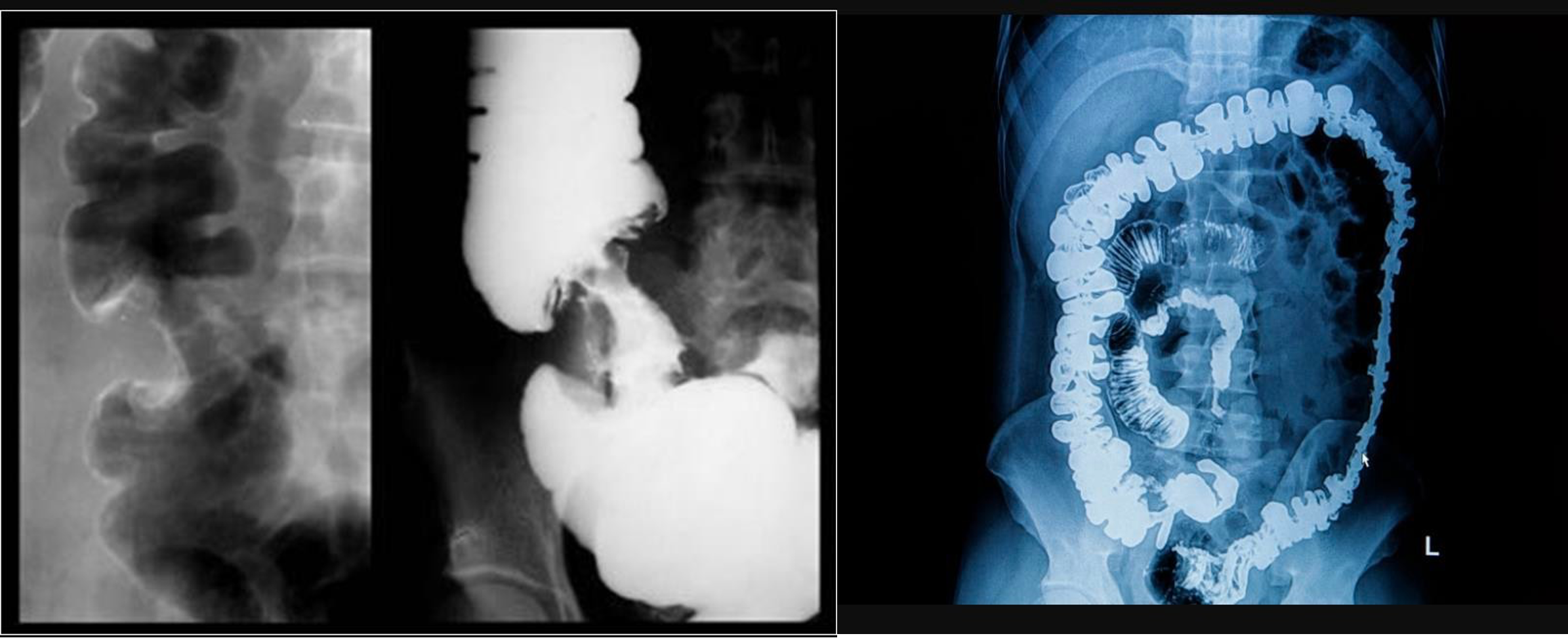
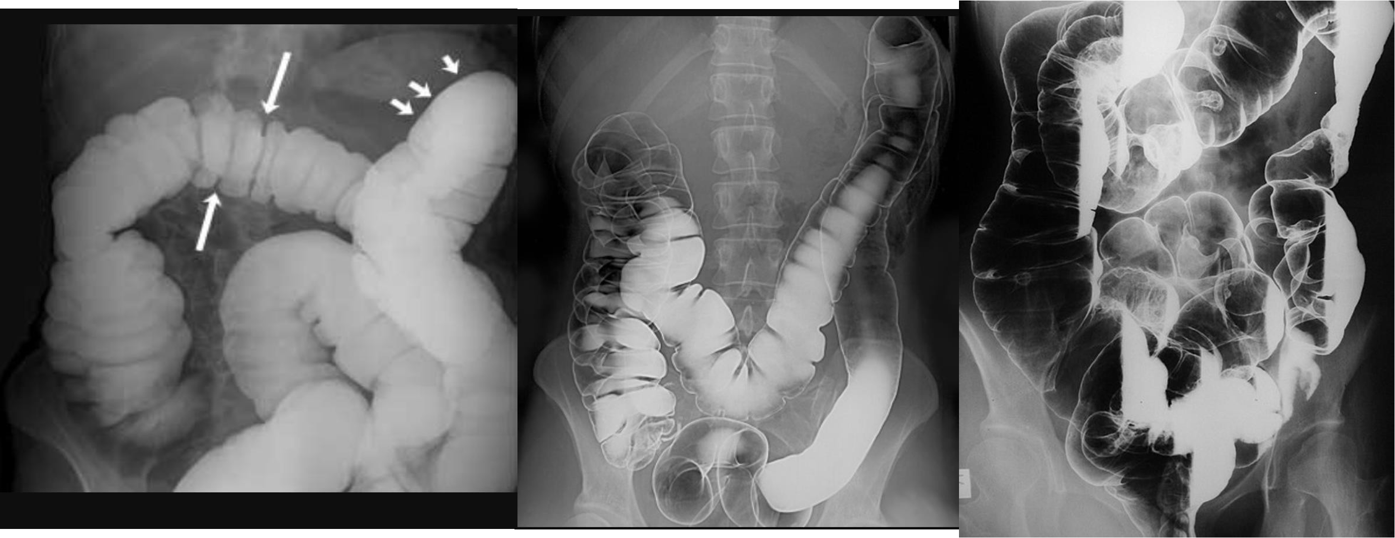
- barium enema from below
- apple core - neck and shoulder - stricture (cancer)
- Ulcerative collitis loss of haustration
- double enema - polyps
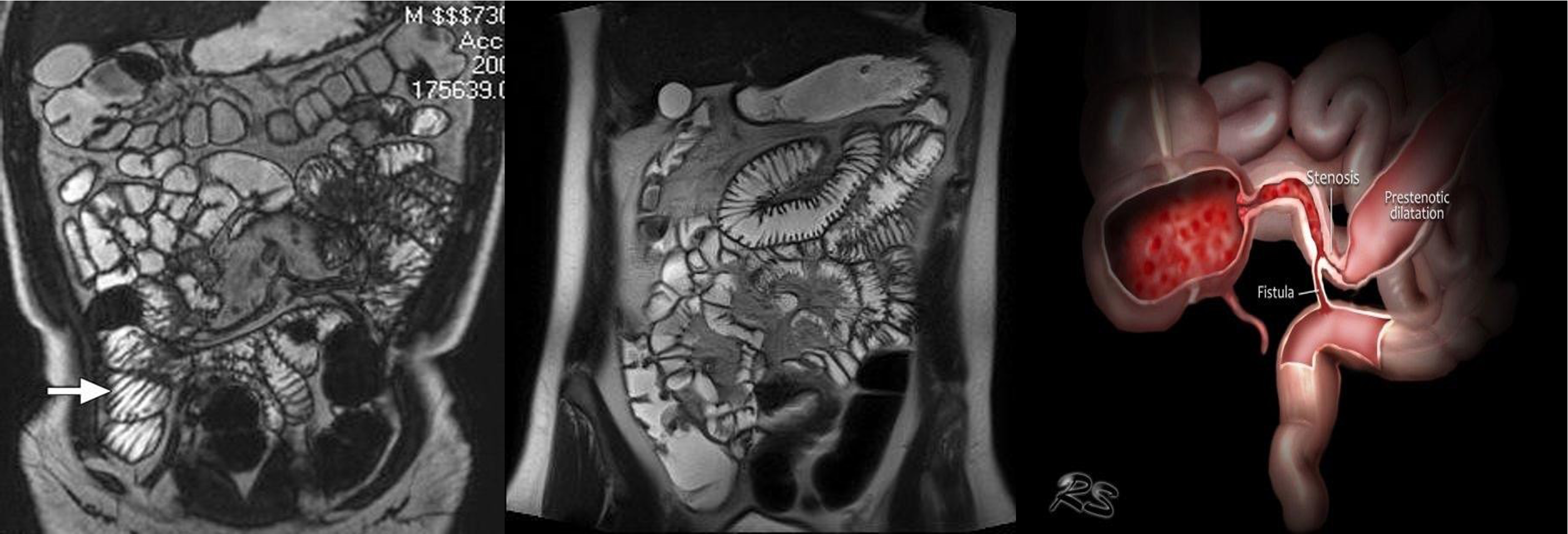
- & 2 MRE -
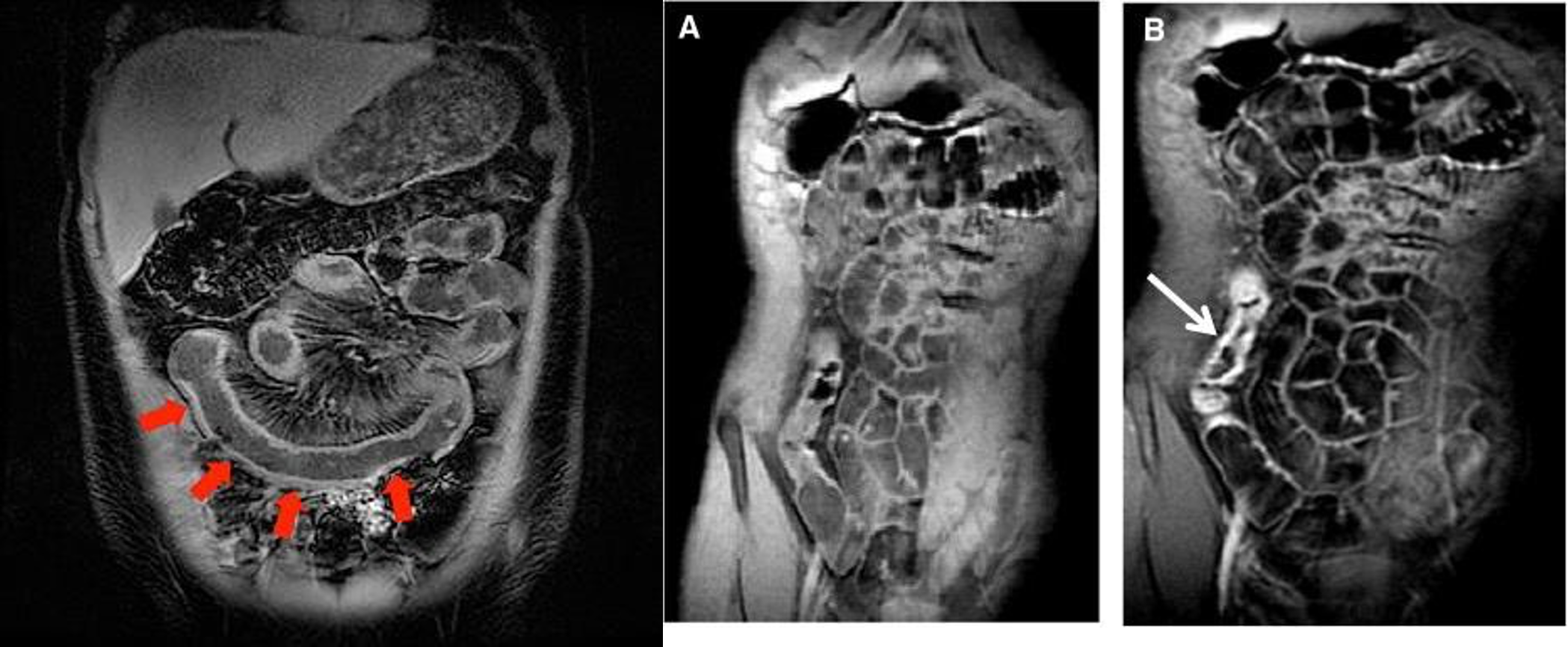
- MRE (crohn)
- MRE
- MRE
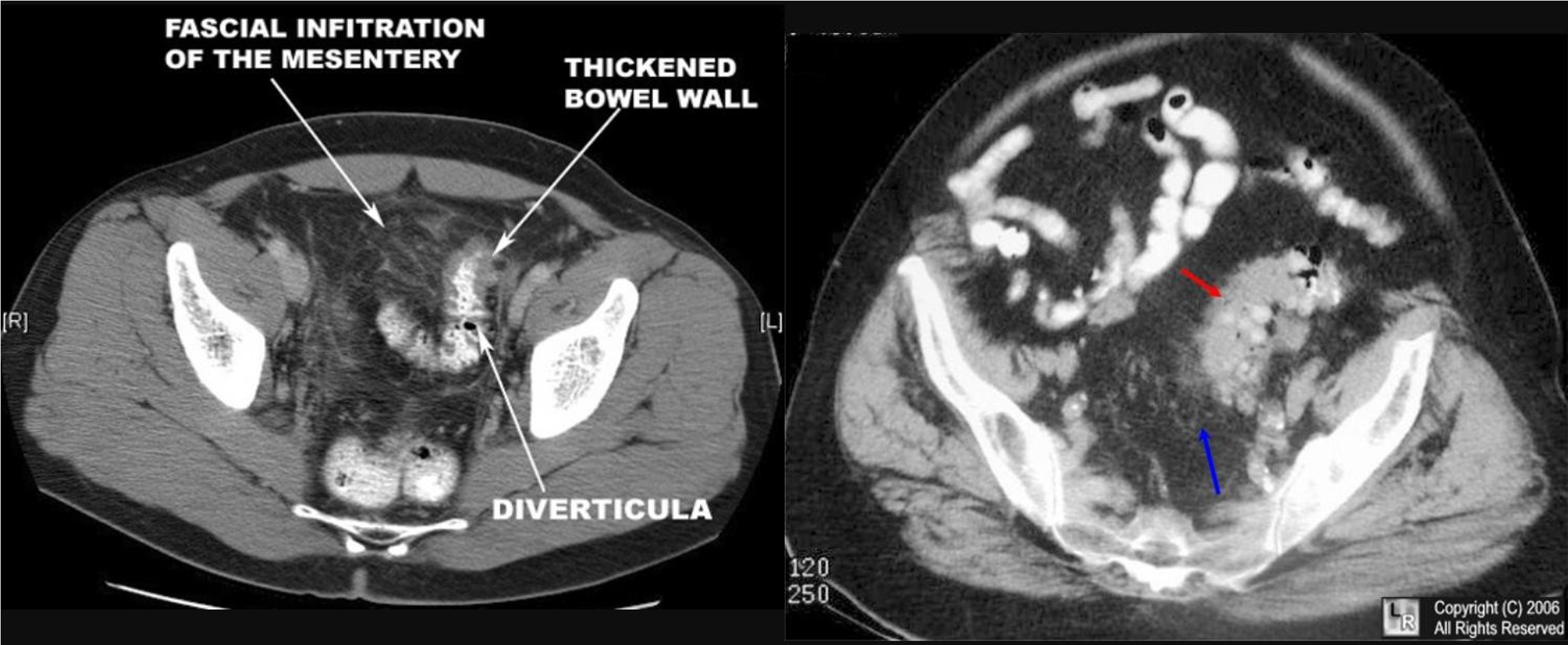
CT
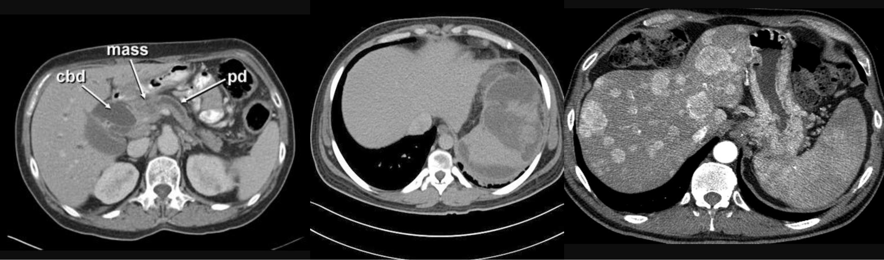
- CBD + METASTESIS
- hypodense - spleenomegaly with splenic infarct
- patient compl anemia vomit - mass stomach - adenocarcinoma - stage 4 gastric cancer (cannon ball sign) - ct scan contrast porta
-
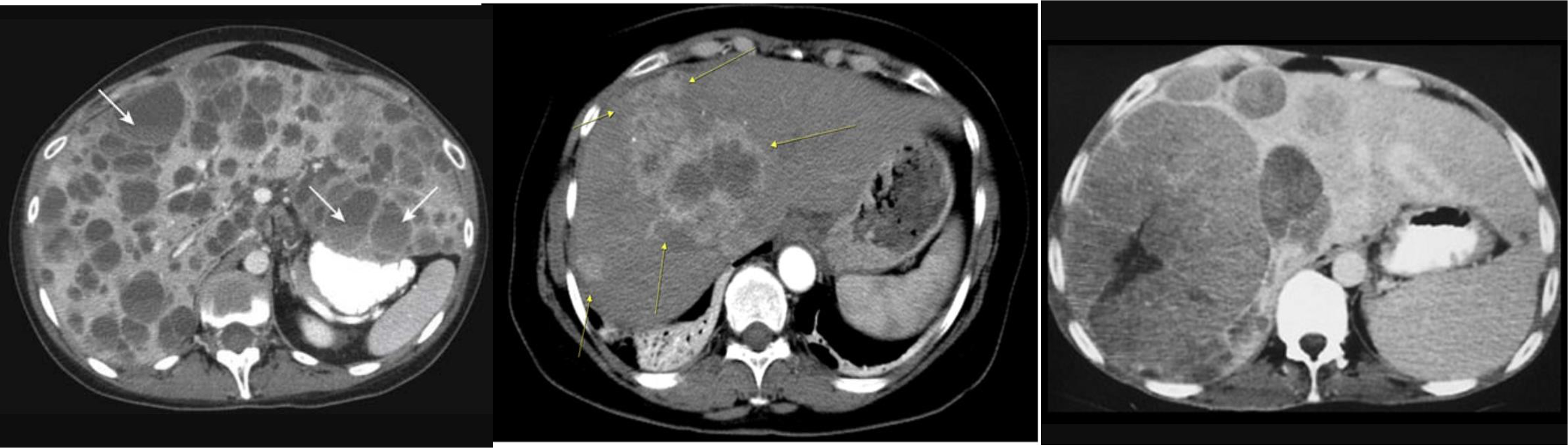
multiple lesion in liver due (Diff; cancer, multiple abcess pyogenic most likely)
-
Ireggular lesion peripheral enhancement - hypo in cent - febrile tender (liver abcess) - antibiotic + ct u/s guidance drainage
-
HCC, Cancer of liver, hepatic adenoma, FNH (with central scar star like)
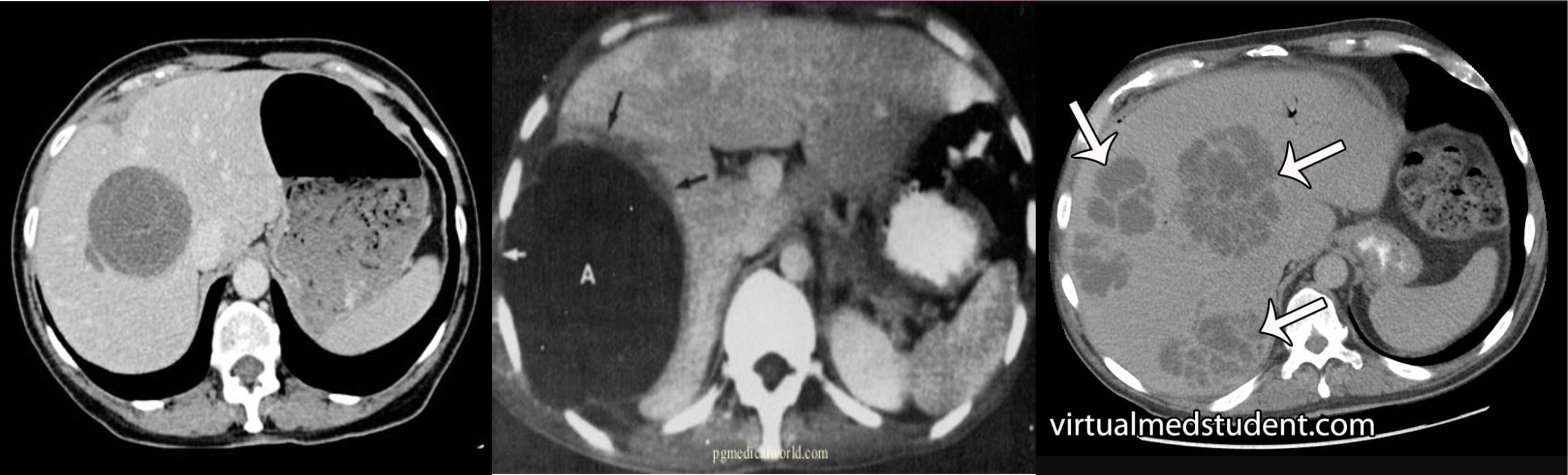
- ct scan (hypodense lesion in liver well circumscribed single - most likely amoebic liver abcess)
- multiple … metastasis
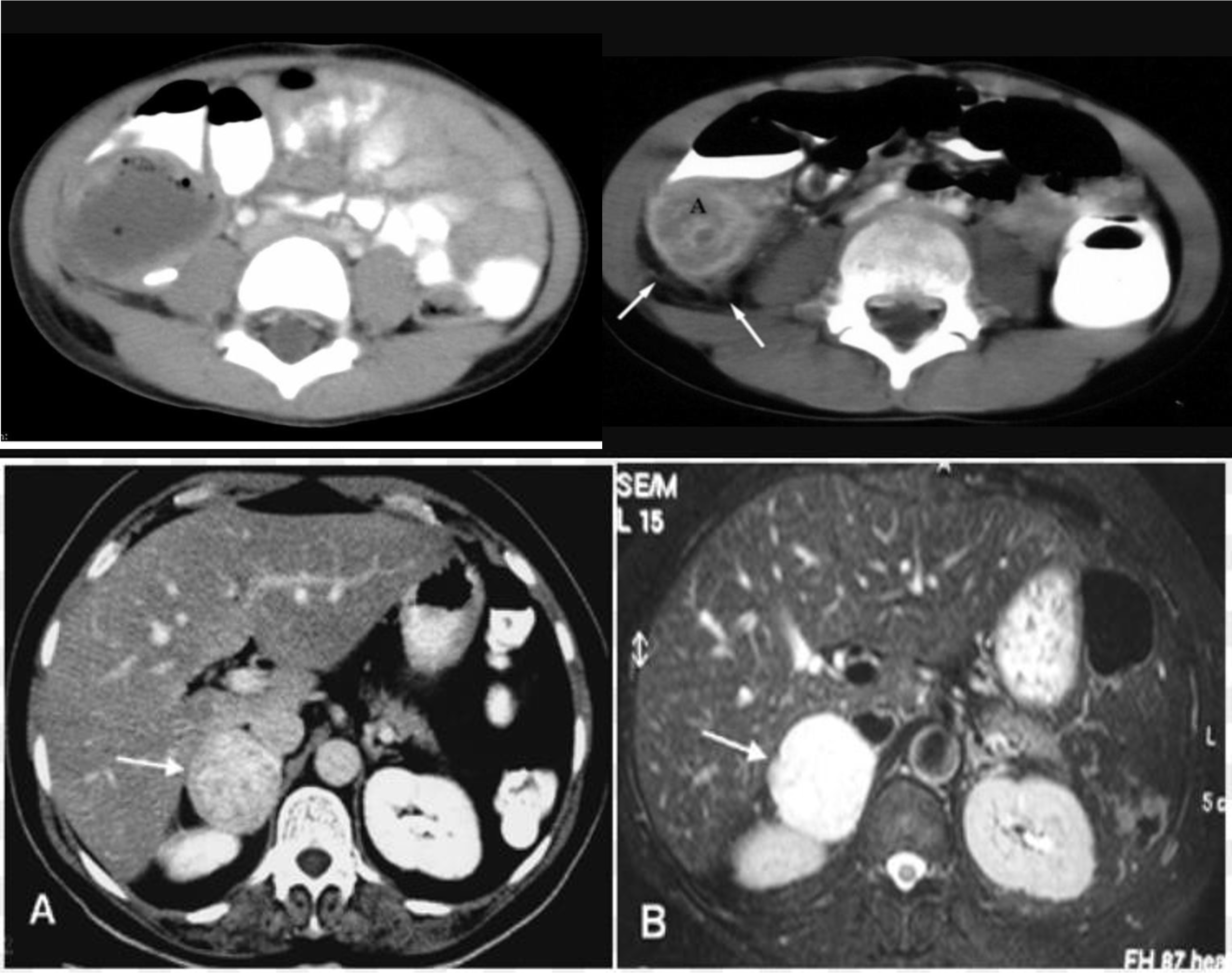
- fullness tenderness right illiac fossa 5D hx - appendicular mass incisional and drainage with antibiotic
- abscess
- ////
- ////
- ////
- ////
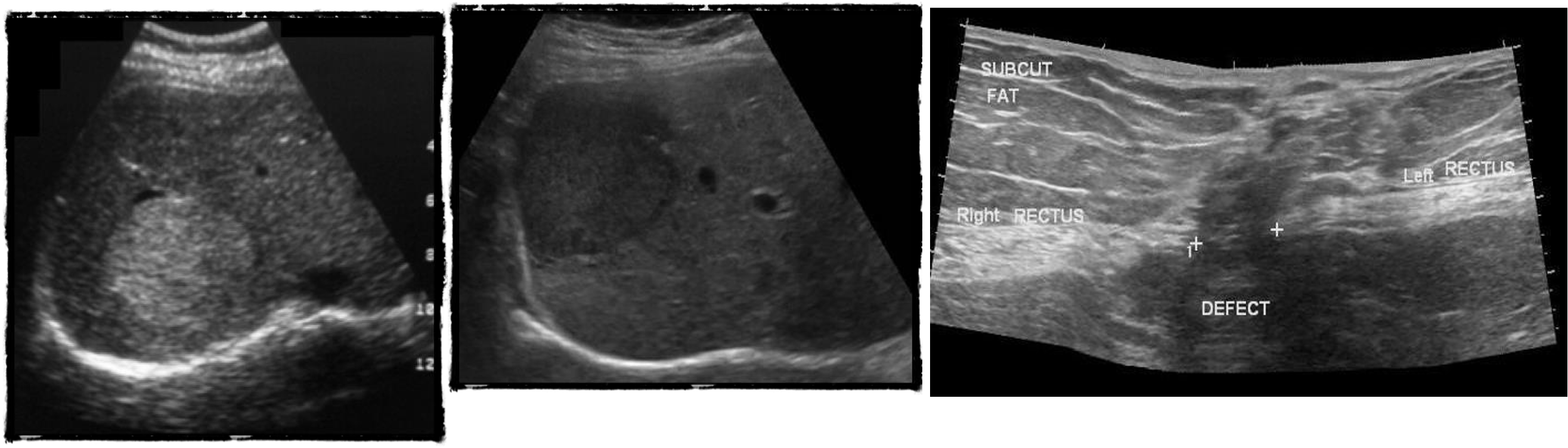
U/S
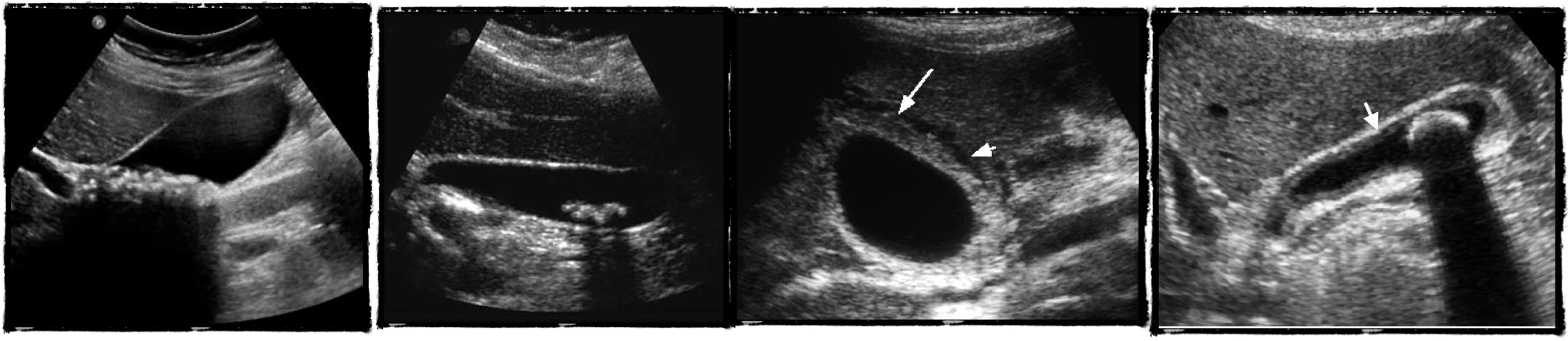
- chronic chole
- hyperechoic - post acoustic shadow…
- acute cholecystitis
- acute edematous thick gallbladder
- stone tumour
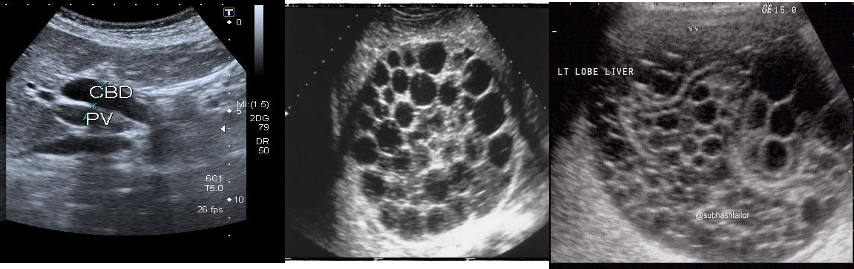
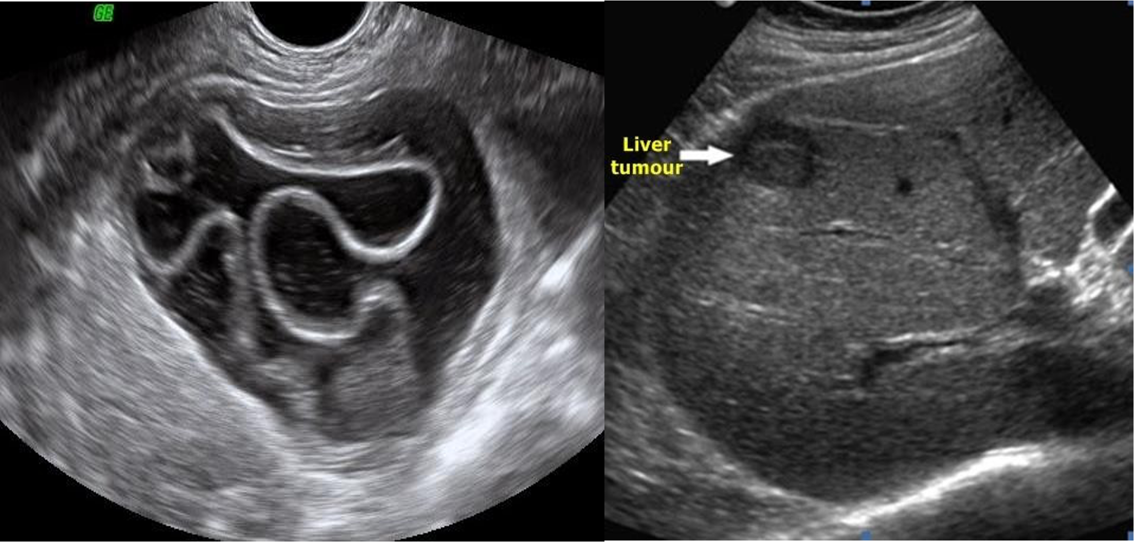 hydiatted
HCC
hydiatted
HCC
 ///
///
///
///
///
///
ERCP
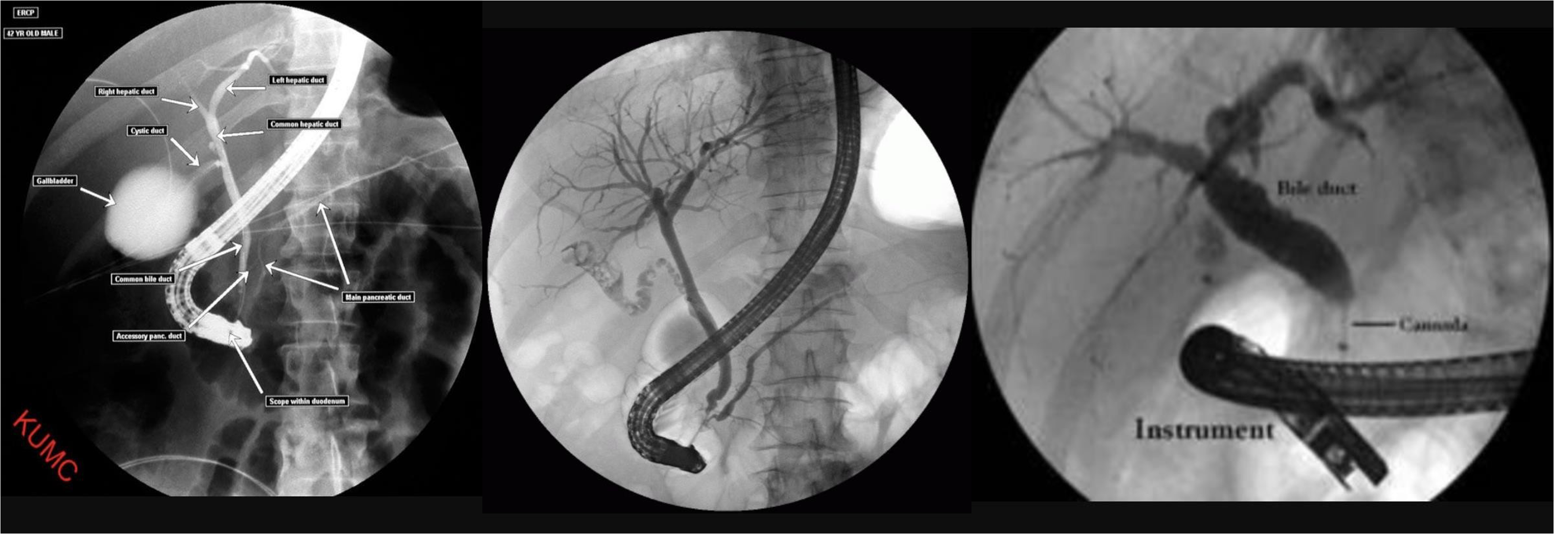
- ERCP
- ERCP
- ERCP dilated (diff stone, tumour)
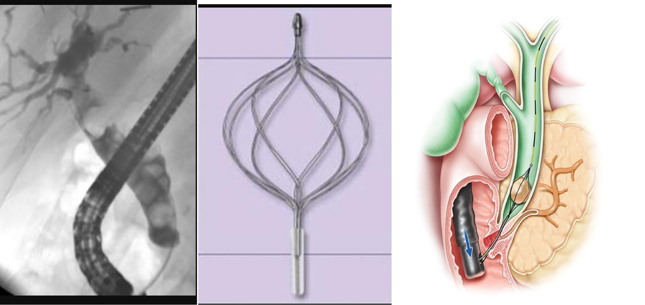
- dilated cbd, stone
- 3 - d. basket?
HIDA SCAN
gall - 100% - diff acute cholecystitis
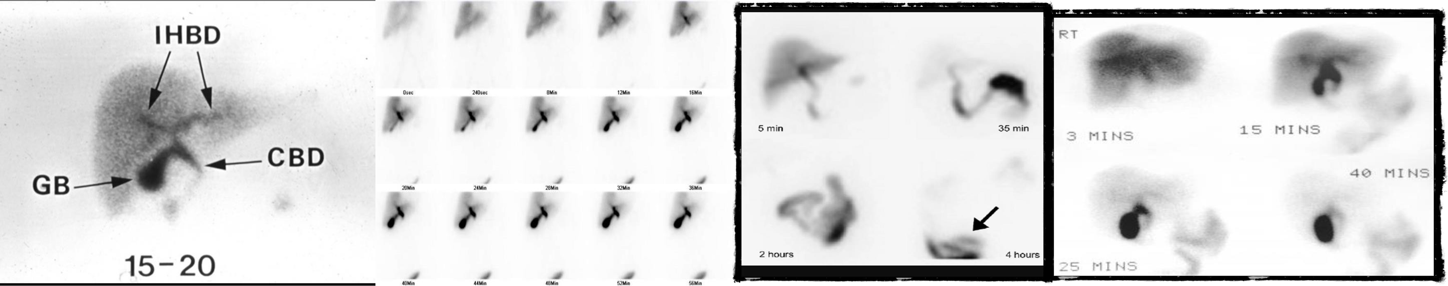 2) negative
3) positive
4)
2) negative
3) positive
4)
MRCP
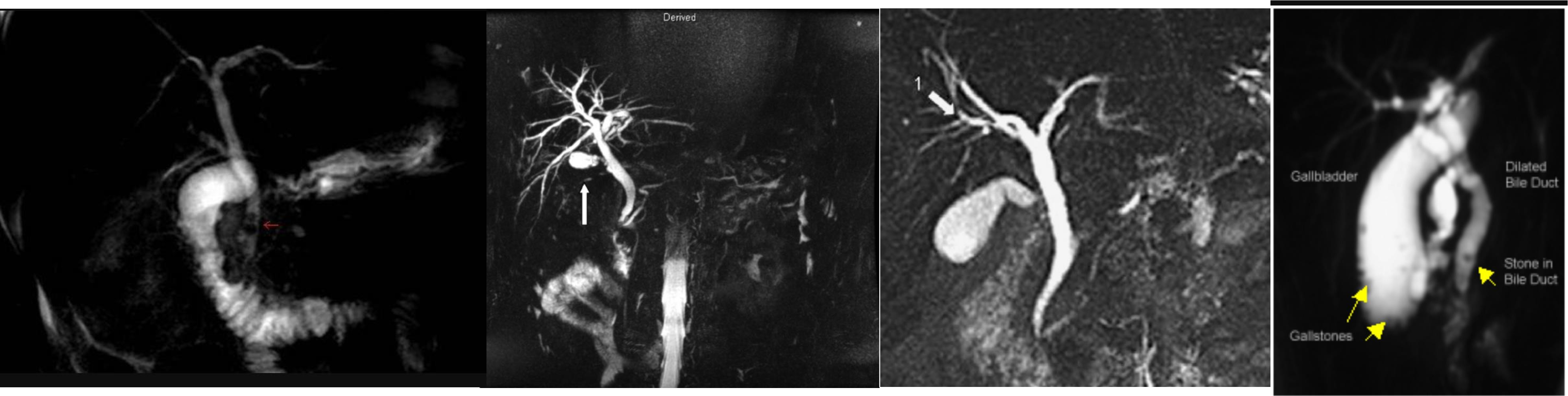 diagnostic therapeutic anatomical
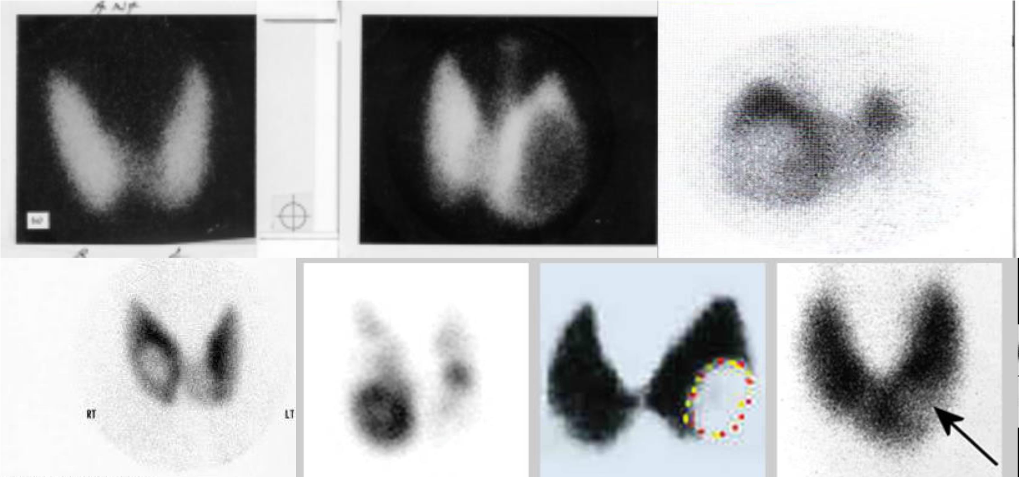
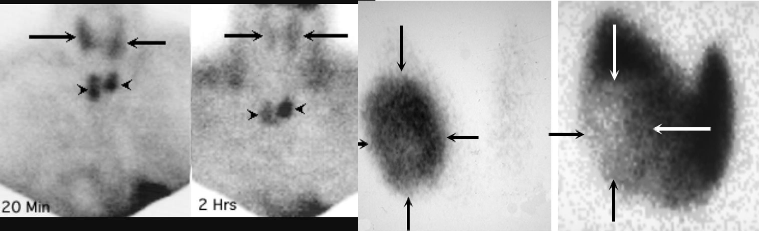
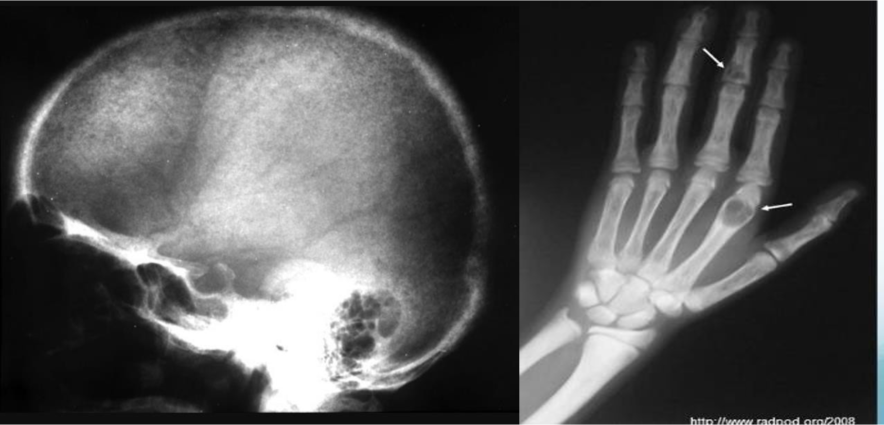
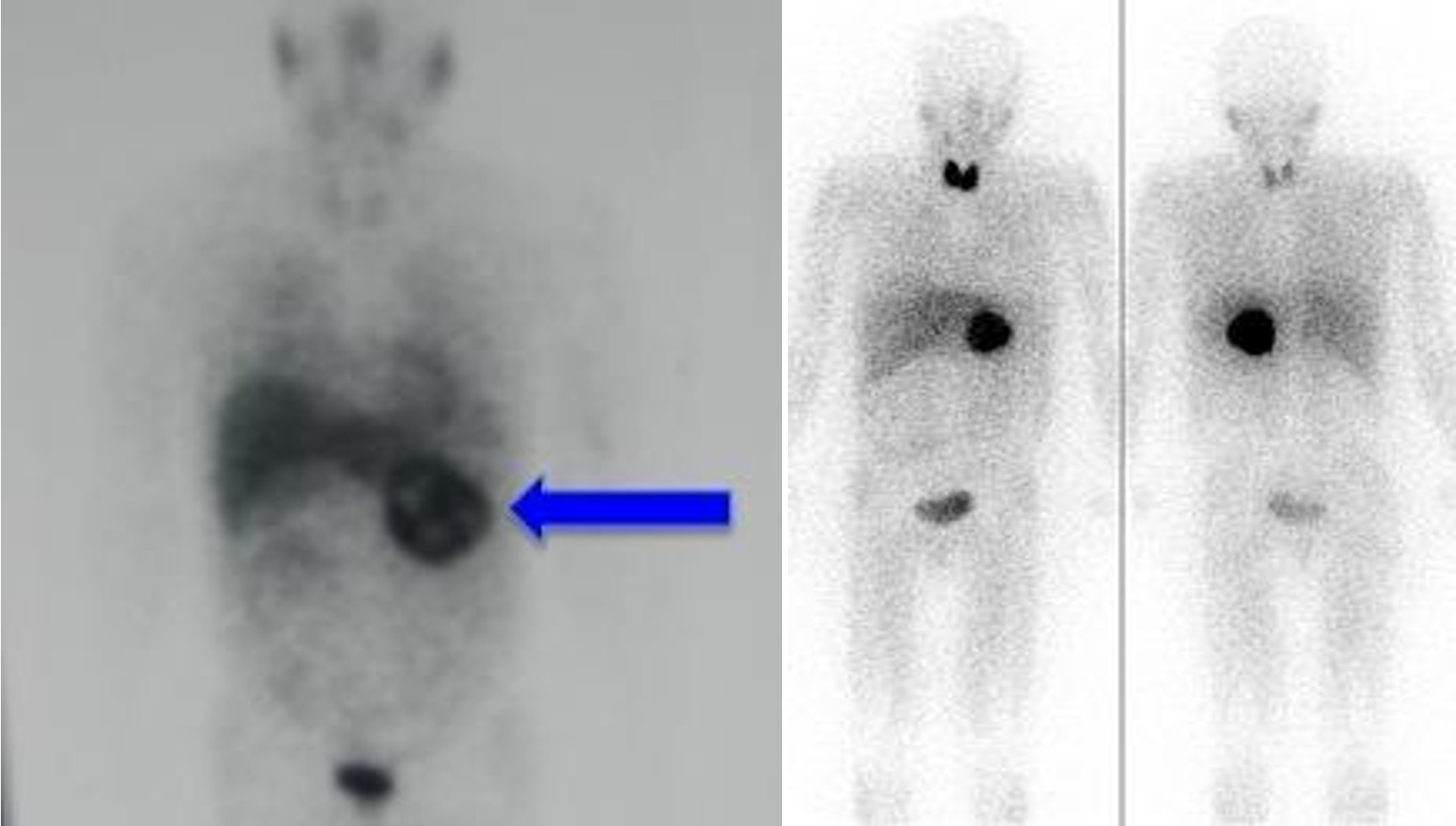
NUCLEAR SCAN
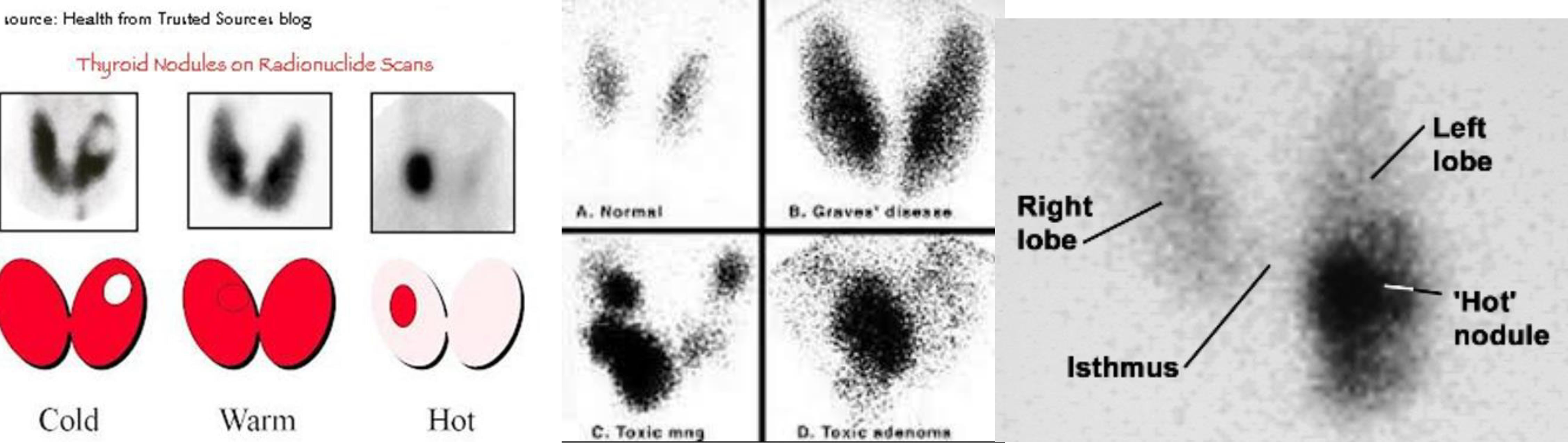
cold nodule = malignancy
MAMMOGRAM
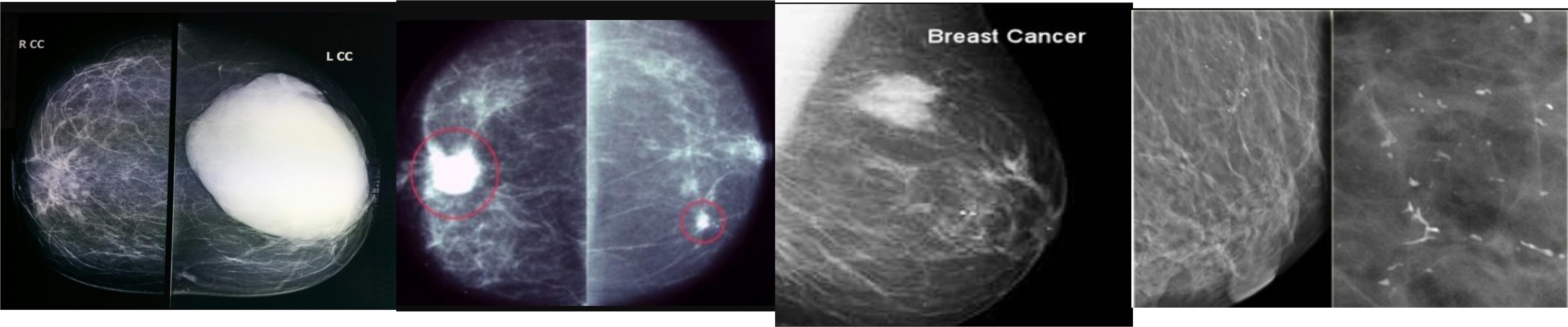 fibroadenoma
PET SCAN
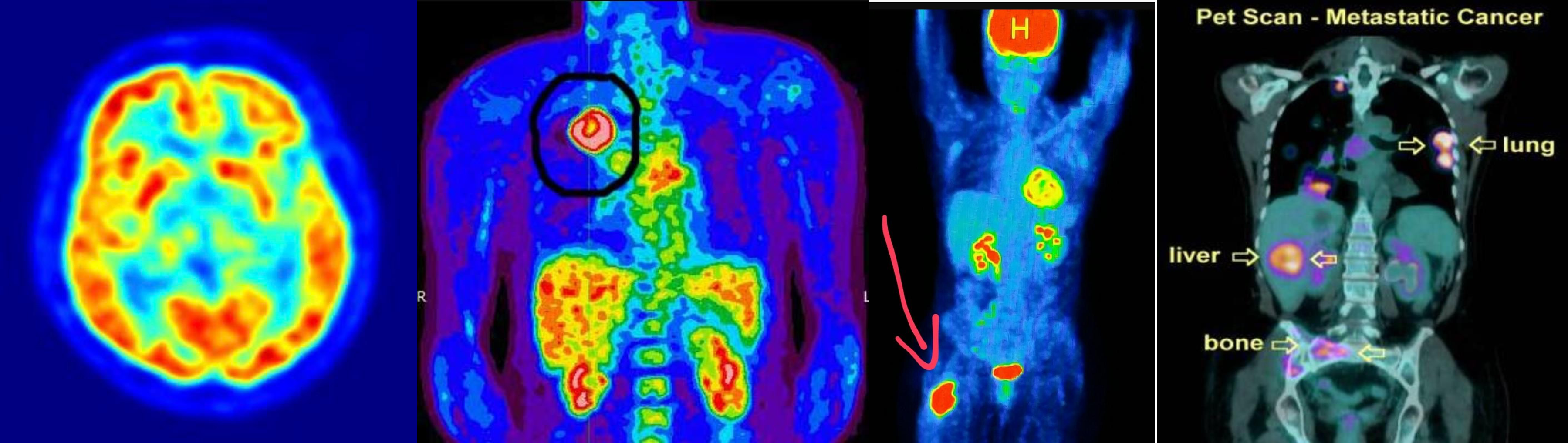 annual follow up
- soft tissue sarcoma -
- lung cancer metasteseses to bone