Principles of Fractures
Prof. Mamoun Kremli
Objectives
- What is a Fracture - the soft tissue part
- Fracture types / classification
- Relation between fracture and force
- History and physical exam. In fractures
- Principles of imaging
What is a fracture?
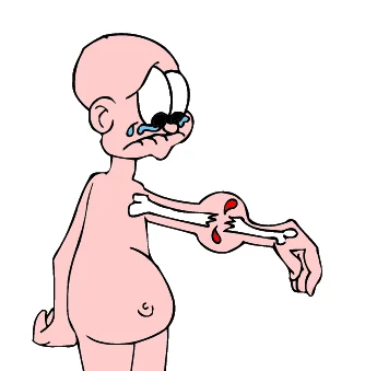
- A fracture is a break in the structural continuity of bone
Definition of a Fracture
- A fracture is a break in the structural Discontinuity of bone
- Always associated with some soft tissue injury Fibula is always lateral
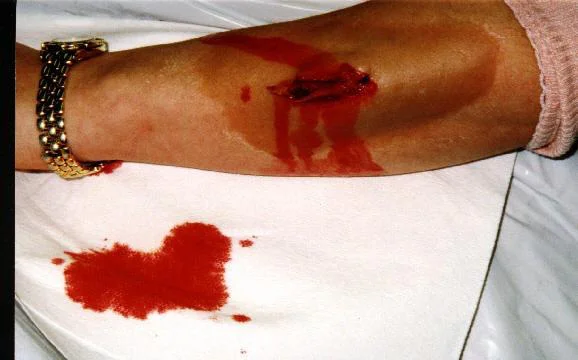
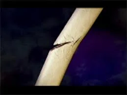
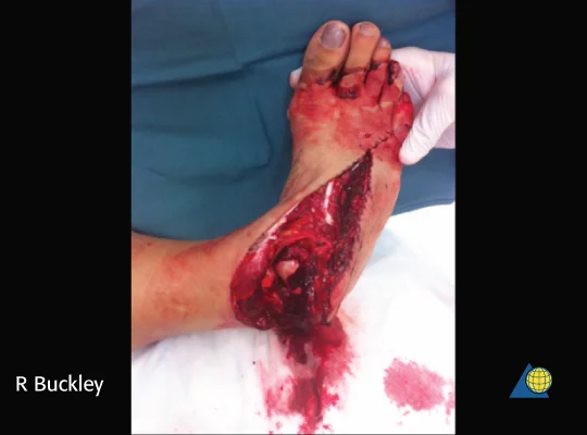
- A fracture is a soft tissue injury in which the underlying bone is broken!
Type of Injury
- Mechanism of injury helps expect the
- Extent and type of bone injury
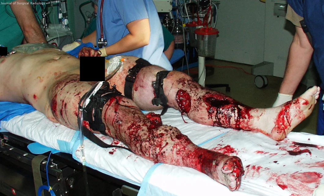
- Simple / comminuted / complex
- Associated fractures/injuries
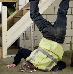
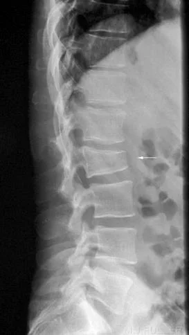
- Fall from height on feet
- fractured calcaneus and lumbar spine
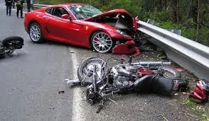
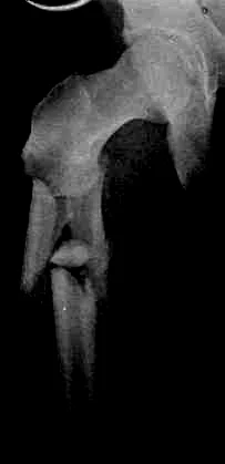
- Car dashboard injuries
- fractured patella and hip dislocation
- Fall from height on feet
- Extent of soft tissue injury
- Suggested treatment and reduction technique
- Prognosis
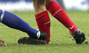
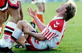
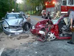
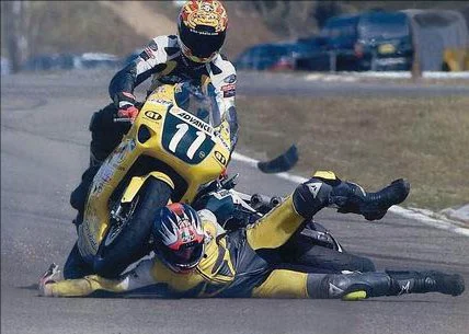
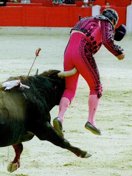
Types of Injury Mechanisms
- Fall: height, point of impact, twist
- Sport: type, direction of force
- Road traffic accident (RTA):
- Car (MVA), motorcycle, pedestrian
- Heavy object fall:
- TV, wall, metal, earthquake
- Assault & firearms / blast


Mechanism of Injury
- Low energy
- High energy
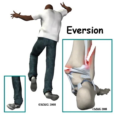
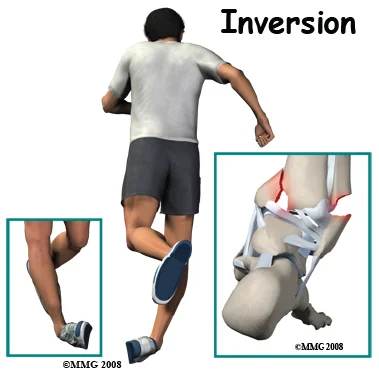
- Direction of force
- Inversion
- Eversion
- Blunt / Sharp
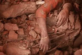
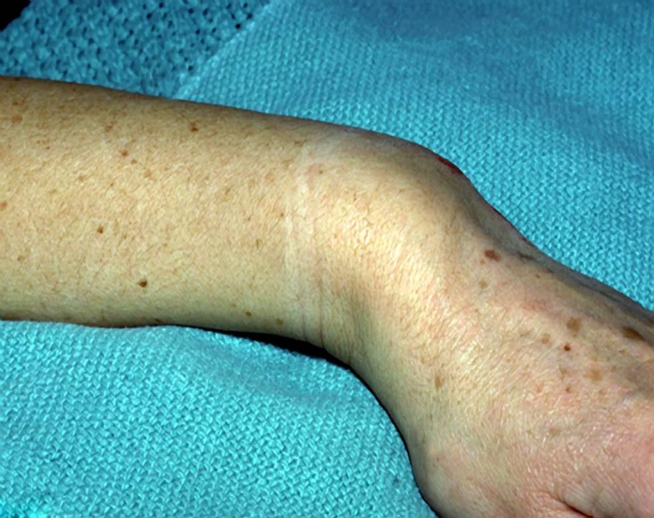
- Closed / Open When the hematoma is exposed to the external environment

Energy Dissipated During Injury
Kinetic Energy = ½ MV²
- If a Simple fall = 1
- Skiing injury = 3-5
- High-velocity gunshot = 20
- Car bumper (25 km/hr) = 100


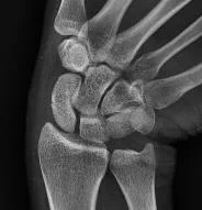
Fracture Classification
- According to site of Fracture:
- Diaphyseal
- Metaphyseal
- Articular
- Epiphyseal (in children)

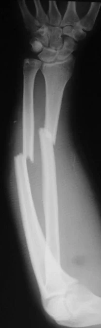
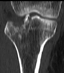
Classification by Fracture Line
- According to fracture line:
-
Complete (usual)
- Cortex fractured on both sides
-
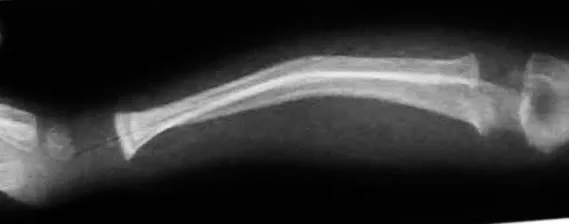
Incomplete (in children)
- Green stick / Torus, Buckle /Deformation
-
Classification by Fracture Pattern
- According to fracture pattern:
- Simple
- Wedge comminuted
- Complex comminuted
- Multi-fragmented

- According to fracture pattern:
-
Compressed
-
Depressed
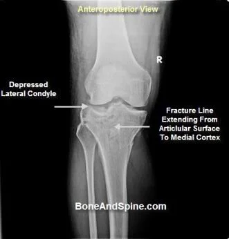

-
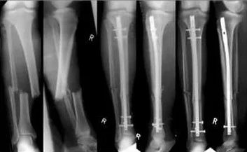
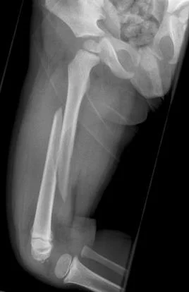
Classification by Type of Injury (Force)
- According to type of injury (force):
- Ordinary fracture
- Expected from force of injury
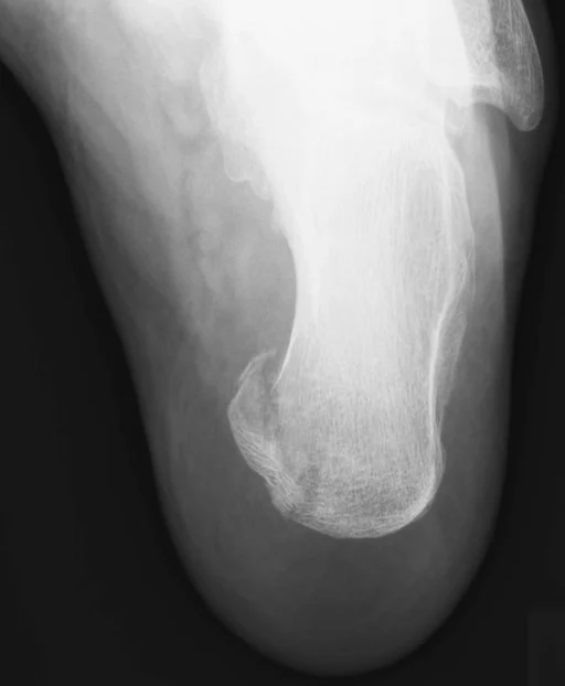
- Stress fracture
- Repetitive loading
- Pathological fracture
- Force too weak to cause fracture
- Bone is pathologically weak
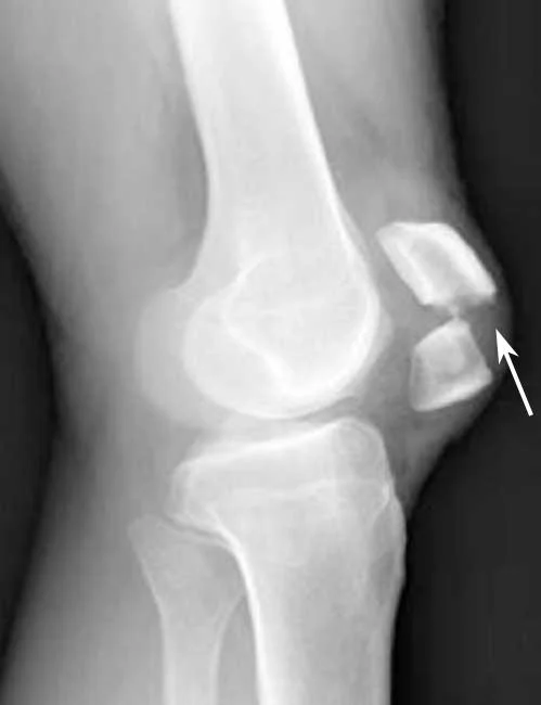
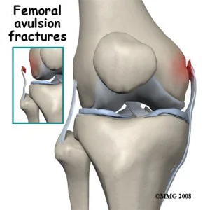
- Avulsion fracture
- Resisted muscle action, or where ligaments and tendons pull a bone fragment off
- Ordinary fracture
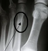
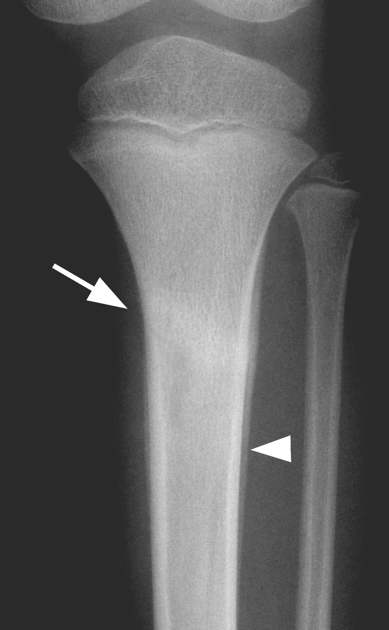
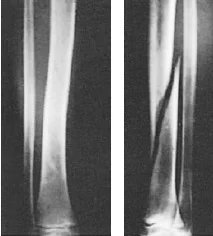
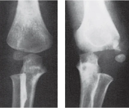
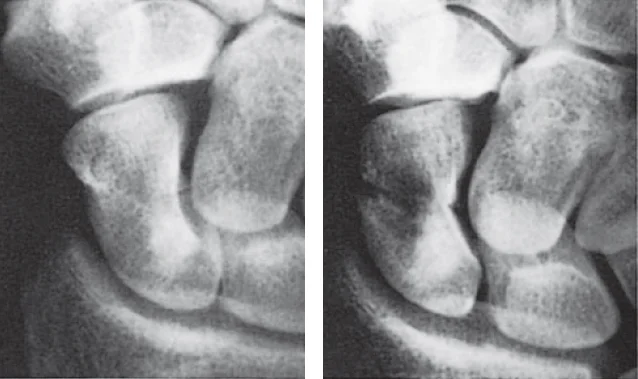
Stress Fractures
- Bone reacts to repeated loading, may become fatigued & a crack develops
- Fatigue fractures
- Abnormal stress or torque on a bone that has normal elastic resistance
- Examples:
- military recruits, athletes, ballet dancers
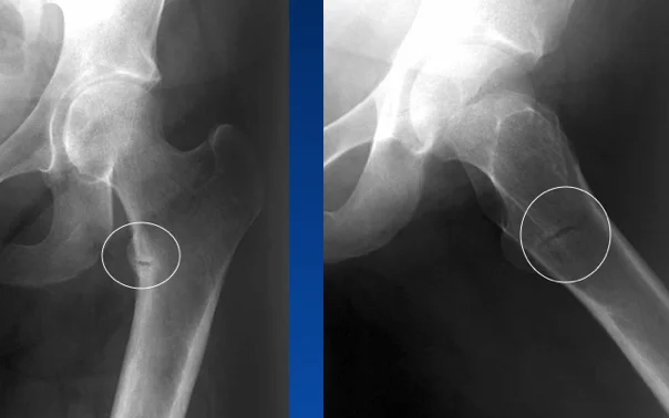
- Insufficiency fractures
- Normal muscular activity stresses a bone that is deficient in mineral or elastic resistance
Stress Fracture Details
- Fatigue fractures
- Usually Transvers
- 2nd metatarsal
- Tibia
- Fibula
- Usually Transvers
- Insufficiency fractures
- In osteopenia, osteomalacia
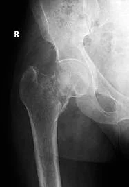
- Neck of femur
- Ribs
- Neck of humerus
- Scapula
- In osteopenia, osteomalacia
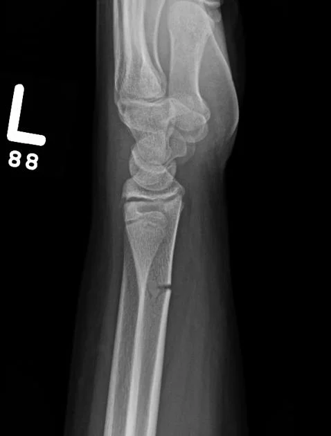
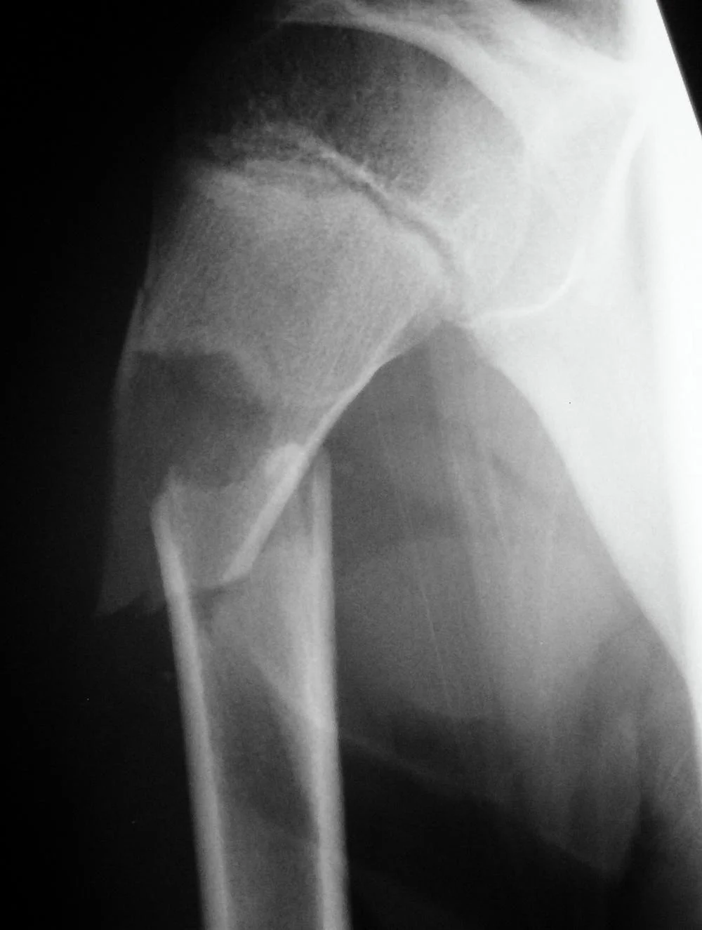
Pathological Fractures
- Fractures caused by trivial force on abnormally weak bone. Seen in:
- Local bone disease
- Osteomyelitis
- Benign tumors and Bone cysts
- Malignant tumors and metastasis
- Generalized disease
- Metabolic: osteoporosis, rickets
- Congenital: osteogenesis imperfecta
- Others: Paget’s disease
- Local bone disease
Force & Fractures
-
Normal bone:
- Strong force: ordinary fracture
- Repetitive stress: Stress (fatigue) fracture
-
Weak bone (Pathological fracture)
- Weak (trivial) force: pathological fracture
- Normal daily activity: Insufficiency fracture
| Quality of Bone | Type of Force | Type of Fracture |
|---|---|---|
| Normal | Strong | Normal |
| Normal | Repetitive loading | Stress - fatigue |
| Abnormal - weak | Normal daily activities | Stress - insufficiency |
| Abnormal - weak | Trivial injury | Pathological |
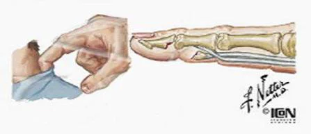
Avulsion Fractures
- Part of bone separated by forceful sudden resisted muscle action
- Caused by ligament or tendon pull on bone
- Part of bone avulsed – bone weaker than tendon/ligament

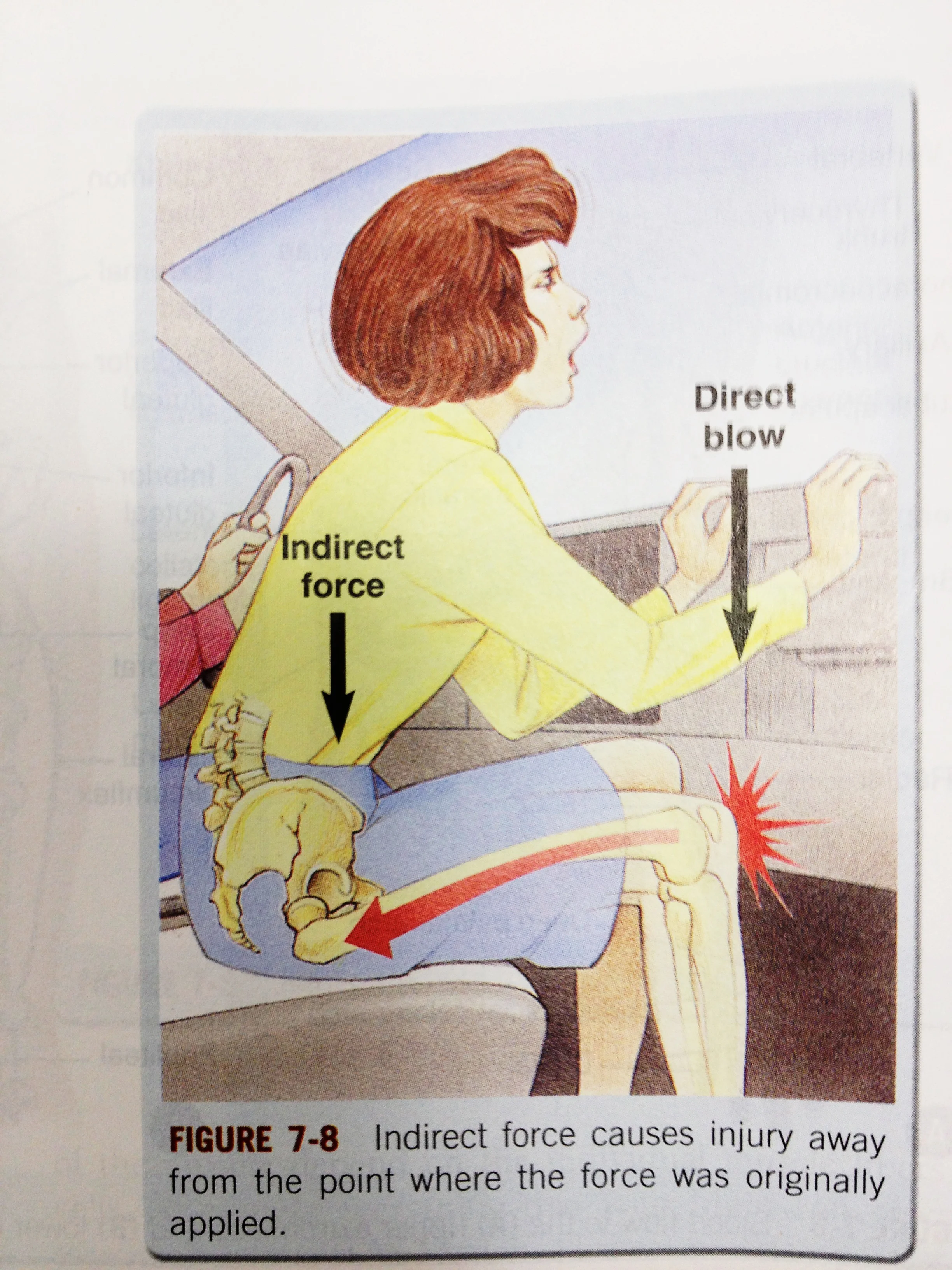
Type of Injury and Fracture Pattern
-
Direct
- Mild force: transverse / Severe force: comminution
- Soft tissue more injured
-
Indirect
- Pattern of fracture depends on force direction
- Less soft tissue injury
-
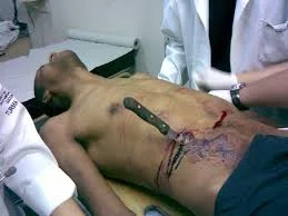
Penetrating
- Missiles
- Low velocity < 300 m/s - damage along the tract
- Comminution
- High velocity: >300m/s - severe comminution
- Comminution with wide soft tissue damage
- Low velocity < 300 m/s - damage along the tract
- Missiles
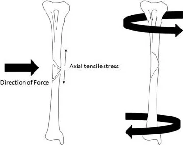
Fracture Pattern and Mechanism of Force
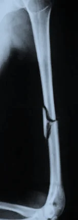
- Fracture pattern suggests mechanism of force
- Spiral: (twisting)
- Short oblique: (compression)
- Wedge: (compression + bending)
- Transverse: (angulation) (avulsion)


Displacement
Types of Displacement
- Described as: Position of distal in relation to proximal
- Un-displaced
- Shift
- Sideways
- Shortening
- Distraction
- Angulation
- In all planes
- Rotation


Fracture Diagnosis
- History
- Clinical features
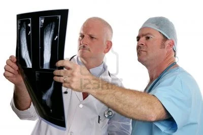
- Imaging: Radiology (x-Ray)
Trauma History
- Mechanism of injury
- Date, time, type, method of impact, …
- Consciousness
- Function of injured part
- Open wound / bleeding
- Other injuries
- Anti-Tetanus status (if skin breached)
Approach - History
- Details of injury
- Mechanism, force, bleeding, consciousness, …
- Details of facture
- Deformity, pain, loss of function, ..
- Other medical problems
- Shall be discussed separately
- Anti-tetanus status if open injuries
- Careful:
- Fractures are not always at the site of impact
- Some fractures do not need severe force
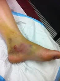
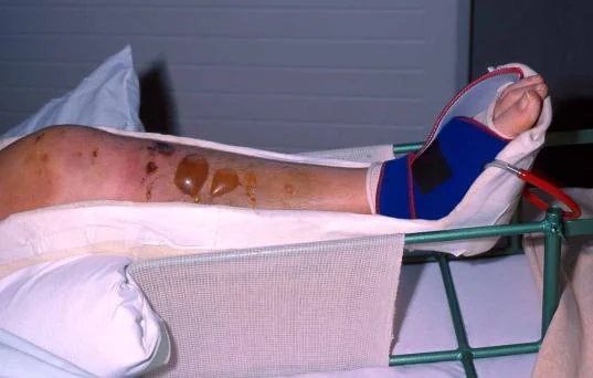
Clinical Features
- History of Trauma
- Symptoms and signs:
- Pain
- Swelling
- Deformity
- Loss of function
- Localised bony tenderness
- Loss of motion
- Abnormal movement
- Crepitus
Approach - Clinical Exam
- General medical condition
- Should be evaluated to exclude
- Shock
- Brain injury
- Other Principles
- Should be evaluated to exclude
- Vital signs
- Should be observed and followed up
-
Look:
- Adequate exposure
- General on patient
- Local:
- Swelling, deformity, bruises, color, …
- Special attention is to be paid to wounds
-
Feel:
- Localized bone Tenderness
- Pulse distal to injury – capillary refill
- Sensory and motor deficits
- Compartment syndrome
- Temperature and crepitus on movement
-
Move:
- With care
- make sure not to cause more pain or injury
- Crepitus & abnormal movement indicates a fracture
- Joints distal to the affected area
- With care
Examination of Viscera
- Examination of the viscera
- Liver and spleen in rib fractures
- Urinary bladder and urethra in pelvic fractures
- Neurological examination in head and spinal injury
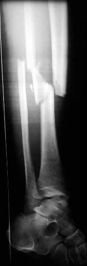
Imaging Principles
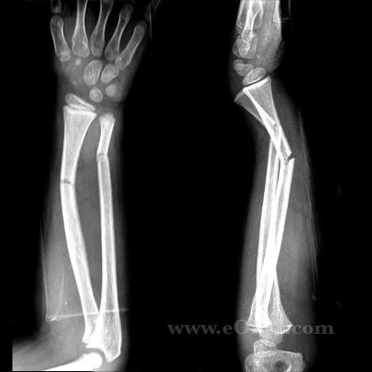
- Plain x-ray: (law of two’s)
- Two views: AP and Lateral
Apley’s System of Orthopedics & Fractures

Law of Two’s
- Plain x-ray: (law of two s)
- Two views: AP and Lateral
- Two joints: joint above and joint below
- To show other injuries
- To assess rotation
- Two limbs: for comparison
- more in children to compare epiphysis
- Two occasions
- e.g. stress fractures
- e.g. scaphoid fracture
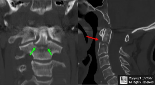
- Two injuries
- e.g. patellar fracture and hip injury
- e.g. calcaneal fractures & spine injuries
- …and two Doctors!!
- Special views:
- Ankle mortis
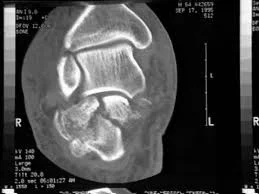
- Calcaneal view
- Scaphoid views
- Shoulder dislocation: axial view
- Acetabular fractures: 45° tilt views
- Stress views
- Traction views
- Functional flexion/extension (spine)
Apley’s System of Orthopedics & Fractures

Advanced Imaging
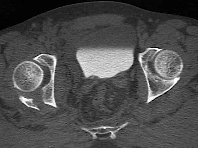
- CT Scan:
- In complex and intra-articular fractures
- In spine
- In pelvic and acetabular fractures
- In calcaneal fractures
Summary
- What is a Fracture - the soft tissue part
- Fracture types - classification
- Relation between fracture and force
- Principles of imaging - Law of “Two’s