Surgery
Necrotizing fasciitis:
Soft tissue infection, in which there is destruction and necrosis of tissue in skin, subcutaneous tissue, muscles. Usually follows abdominal surgery or trauma.
Caused by:
- Infection with group A Streptococcus, commonly known as “flesh-eating bacteria”
Others include:
- Aeromonas hydrophila
- Clostridium
- E. coli
- Klebsiella
- Staphylococcus aureus
Immuno - compromised [e.g. Diabetics and IV drug addicts] are more susceptible.
Common sites: abdominal wall, perineum and limbs.
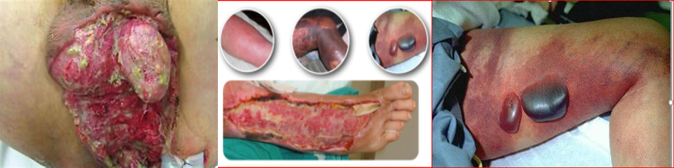
Starts as cellulitis with early systemic toxicity. It is characterized by non-blanching erythema, with blisters and frank necrosis of the skin. No definitive margins
TREATMENT:
Extensive surgical debridement of the affected area, in combination with high dose broad spectrum is the appropriate Rx till C/S result.
Fourniers gangrene: is defined as a polymicrobial necrotizing fasciitis of the perineal, perianal, or genital areas
Dermatology
Necrotizing Fasciitis
- Necrotizing fasciitis is a life-threatening infection of the fascia just above the muscle
- Progresses rapidly over the course of hours and may follow surgery or trauma, or have no preceding visible lesion
- Expanding dusky, edematous, red plaque with blue discoloration
- May turn purple and blister
- Anesthesia of the skin of the affected area is a characteristic finding
- Caused by group A streptococcus, Staphylococcus aureus or a variety of other organisms
Treatment
- Considered a medical/surgical emergency with up to a 20% mortality rate
- If suspect necrotizing fasciitis: consult surgery immediately
- Treatment includes widespread debridement and broad-spectrum systemic antibiotics
- Poor prognostic factors include: delay in diagnosis, age>50, diabetes, atherosclerosis, infection involving the trunk

Case Six
History
- HPI: Mr. G is a 66-year-old man who was admitted for an inguinal hernia repair. His surgery went well and he was recovering without complication until he was found to have an expanding red rash on his left thigh. The dermatology service was consulted for evaluation of the rash.
- PMH: hypertension, diabetes mellitus type 2
- Medications: lisinopril, insulin, oxycodone
- Allergies: none
- Family history: noncontributory
- Social history: retired, lives with his wife
- Health-related behaviors: no alcohol, tobacco, or drug use
- ROS: febrile, fatigue, rash is painful
Examination
- Vital signs: T 101.1, HR 110, BP 90/50, RR 18, O2 sat 98%
- General: ill-appearing gentleman lying in bed
- Skin: ill-defined, large erythematous plaque with central patches of dusky blue discoloration, which is anesthetic, upon re-examination 60 minutes later the redness had spread
Question 1
- Which of the following do you recommend for initial management? a. Call an urgent surgery consult b. Give IV fluids and antibiotics c. Image with stat MRI d. Obtain a deep skin biopsy e. All of the above
Case Six, Question 1 - Answer
Answer: e
- Which of the following do you recommend for initial management? a. Call an urgent surgery consult (The suspected diagnosis is a surgical emergency) b. Give IV fluids and antibiotics (Patients quickly become hemodynamically unstable) c. Image with stat MRI (To assess degree of soft tissue involvement. Appropriate, but do not delay surgical intervention) d. Obtain a deep skin biopsy (Helps confirm diagnosis) e. All of the above