orbit
orbit
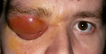
A 35-year-old man presents to his GP with erythematous, swollen right upper and lower eyelids, worsening over the previous 2 days. He is unable to open them. He feels unwell and has a high temperature. History of chronic sinusitis 4 years ago and glasses wearing since 10 years. Examination reveals marked lid swelling, and on manual opening of the lids a proptosis with chemotic injected conjunctiva. Eye movements are limited in all directions. Visual acuity and colour vision are normal, and there is no relative afferent pupillary defect. The optic disc and retina also appear normal.
Diagnosis
Right Orbital Cellulitis
1. Definitions
| Term | Location |
|---|---|
| Preseptal cellulitis | Infection of eyelid/anterior eyelid tissues, anterior to the orbital septum |
| Orbital cellulitis | Infection of orbital fat and ocular muscles, posterior to the septum |
2. Key Clinical Features
| Feature | Preseptal Cellulitis | Orbital Cellulitis |
|---|---|---|
| Location | Infection of eyelid/anterior eyelid tissues, anterior to the orbital septum | Infection of orbital fat and ocular muscles, posterior to the septum |
| Eyelid swelling/erythema | Yes | Yes |
| Tenderness | Mild–moderate | Moderate–severe |
| Ocular pain | Often, but no pain with eye movement | Worse on eye movement |
| Eye movements (motility) | Full range | Restricted |
| Proptosis | No | Yes |
| Vision changes | No | May have ↓ visual acuity / RAPD |
| Systemic signs | May have low-grade fever | Frequently fever, malaise |
| Skin warmth | May feel warm | Often hot to touch |
3. Investigations
-
Clinical exam: carefully assess eye movements, pupillary responses, visual acuity.
-
Imaging
-
CT or MRI orbit with contrast:
-
Confirms posterior (“orbital”) involvement
-
Rules out subperiosteal or intra-orbital abscess
-
Looks for sinus disease, foreign body, tumor
-
-
-
Laboratory
-
CBC (↑ WBC)
-
Blood cultures if febrile
-
4. Management
A. Preseptal (Periorbital) Cellulitis
-
Outpatient (if well-appearing, no systemic signs)
- Oral antibiotics covering Staph/Strep (e.g. cephalexin, dicloxacillin) for 7–10 days
-
Admission & IV antibiotics if:
-
Toxic appearance or high fever
-
Very young child (<1 year)
-
Immunocompromised
-
B. Orbital Cellulitis
-
Hospitalize immediately
-
Empiric IV broad-spectrum antibiotics, covering Gram-positives (including MRSA), Gram-negatives and anaerobes.
- e.g. vancomycin plus a third-generation cephalosporin (ceftriaxone or cefotaxime) ± metronidazole
-
Consider systemic steroids (e.g. dexamethasone) to reduce inflammation and orbital pressure—but only after 24–48 h of effective IV antibiotics and under close monitoring.
-
Surgical drainage if:
-
Subperiosteal or intra-orbital abscess on imaging
-
No clinical improvement within 24–48 h of antibiotics
-
Evidence of compressive optic neuropathy
-
5. “Red Flags” Suggesting Orbital Involvement
-
Proptosis (forward displacement of the globe)
-
Pain or limitation on eye movement
-
↓ Visual acuity, RAPD
-
High fever, systemic toxicity
-
“Hot” (warm) eyelid skin
Key take-home:
-
Preseptal cellulitis → often outpatient, oral antibiotics, good follow-up.
-
Orbital cellulitis → emergency admission, IV antibiotics, imaging, possible surgery.
Diagnosis
Right Orbital cellulitis
Differential diagnosis
- Uveitis
- Sudden painful decrease of vision
- ACG
- Optic neuritis
- Endophthlmitis
- Keratitis
DDx: (red painful eye)
- Endophthlmitis.
- Keratitis.
- Uveitis.
- Glaucoma.
DDx: (unilateral proptosis)
- Trauma.
- Tumor.
- Orbital cellulitis.
- Carotid-cavernous fistula.
- Orbital varix.
- Orbital cyst.
- Orbital varicose.