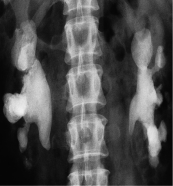
-
Symptomatic/asymptomatic: Flank pain hematuria
-
Most urinary calculi are calcified and show varying density on x-ray examinations.
-
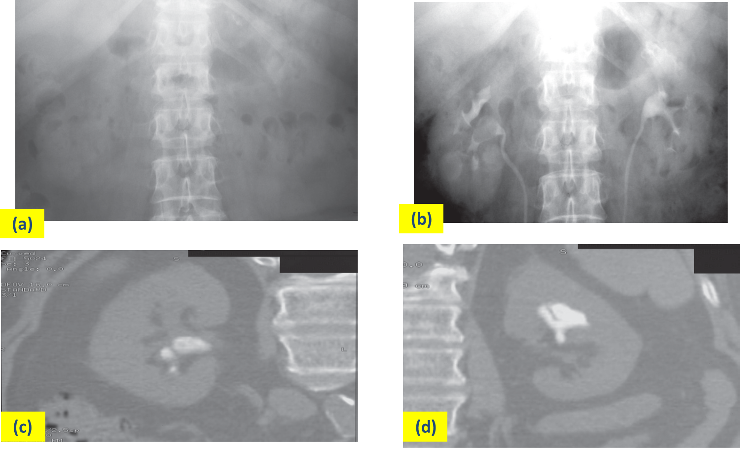
Only pure uric acid and xanthine stones are radiolucent on plain radiography, but they can be identified at CT or ultrasound.
-
All renal calculi have high attenuation on CT

 (a) IVU control film. Renal stones are not visible on the right and are very poorly visualized on the left. (b) IVU following intravenous contrast. Filling defects are seen in the right lower calix and pelvis and in the left upper pole calices . (c, d) CT of the kidneys in the same patient with no contrast medium, reformatted in the coronal plane, demonstrating the renal stones in both the right (c) and left (d) kidneys.