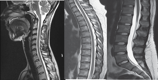
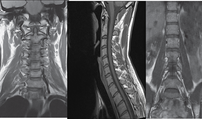
The gold standard of imaging for spinal disorders
-
Can identify abnormalities of bone, discs, muscles, ligaments and spinal cord
-
Intravenous contrast is sometimes administered to better visualize certain structures or abnormalities


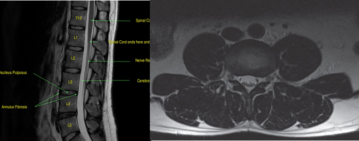
Axial T2-weighted scan through the L3/L4 disc space.
At this level, the L3 nerve roots are in the exit foramina and the L4 nerve roots have moved to the edge of the dural sac in the lateral recesses prior to exiting the spinal canal at the level below.
