Etiology
- High speed car and motorcycle accidents or falls, especially in the elderly
Imaging
Pelvic x-ray: useful for bedside screening, especially in hemodynamically unstable patients
CT abdomen and pelvis with IV contrast
- Gold standard for hemodynamically stable patients
- CT has a higher sensitivity at detecting fractures compared to pelvic X-rays. -
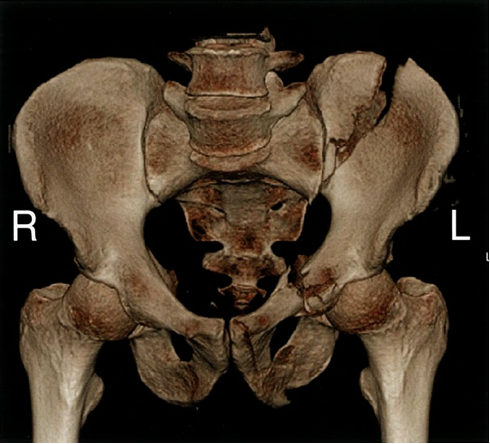
- 3D reconstruction of the bony pelvis can additionally improve surgical planning.
 A. Fractures through the pubic rami (arrows) are common in the elderly.
B. Unstable pelvic ring fracture - X-ray pelvis (AP view) The right hemipelvis is displaced superiorly, compatible with disruption of the sacroiliac joint . Bilateral pubic ramus fractures are also present. - best to assess urethra after incident
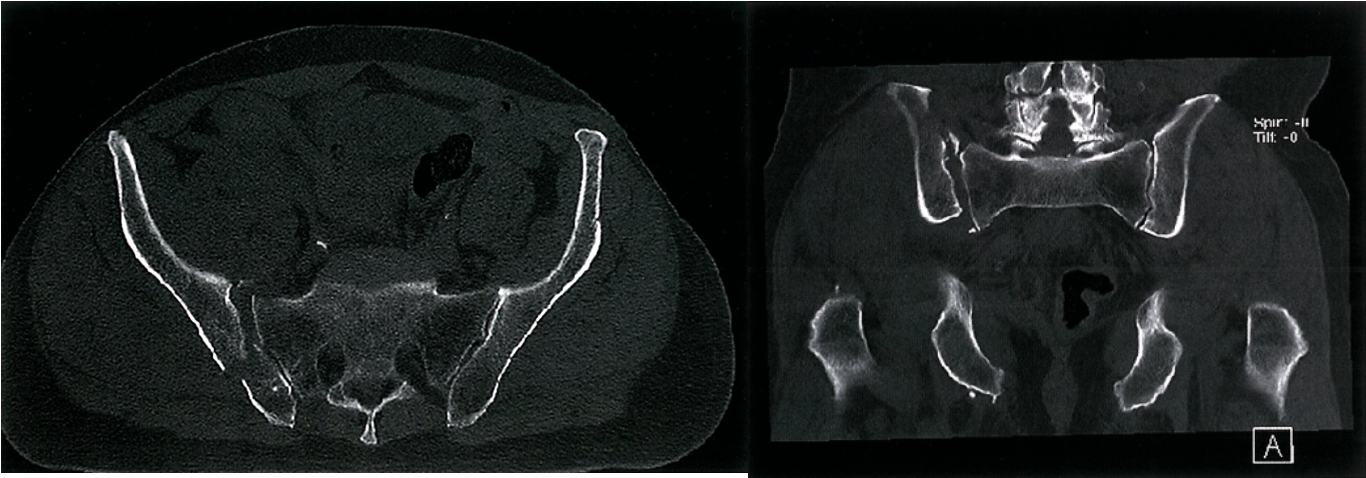
 A. Posterior pelvic ring fracture - CT pelvis (bone window; axial)
There is disruption of the right sacroiliac joint with a fracture demonstrated through the right ilium.
B. Post. pelvic ring fracture - CT pelvis (bone window; coronal) There is widening of the right sacroiliac joint with a small fracture fragment demonstrated at the superior aspect of the joint
 Left sided pelvic fractures - CT pelvis (3D reconstruction)
There is an obliquely orientated comminuted fracture through the left iliac wing communicating with the left sacroiliac joint, as well as fractures of the pubis and the ischium (used mainly for surgical planning)
 Fractures through the femoral neck Z Z
are common in the elderly and may result from minor trauma. The radiographic signs in some instances may be subtle.
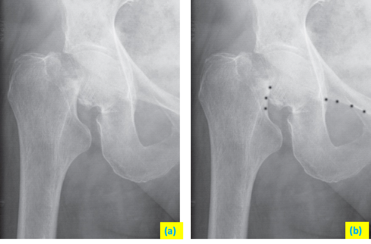
(a) A fracture through the femoral neck interrupts Shenton’s line. z
(b) Shenton’s line Z is a curved line formed by the top of the obturator ring and the medial aspect of the neck of the femur (the interruption of Shenton’s line is shown by the dots).
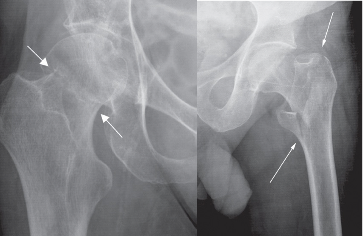
 A. Impacted femoral neck fracture (arrows) causing only a
sclerotic line and disruption of the trabecular architecture. z
B. Intertrochanteric fracture (arrows) between the greater and lesser trochanters.