Medicolegal Identification
Definition
The application of Forensic Science and Technology to personalize someone through specific criteria that individualizes them from others throughout their life. This can be used in living persons, deceased persons, or even parts of a dead body or collections of bones.
Identification of Living Persons
Civil Cases
- Unconscious person admitted to hospital
- Amnesia (memory loss)
- Persons without authentic records when facing legal situations (e.g., marriage)
- Inheritance matters
- Military service eligibility
- Emigration at exit ports
Criminal Cases
- Criminals who impersonate others
- Offenders below age of 18 having no official records of age to be tried in juvenile courts (typically ages 12-18 years)
Main Points of Identification
1. General Features
- Sex, race, age
- Facial features: Color of skin, type and color of hair, color of eyes, moustache, etc.
- Height and weight
2. Clothing (Police Responsibility)
- Check pockets for identity papers
- Note distinctive clothing items
3. Characteristic Marks
- Moles and scars
- Tattoo marks
- Congenital abnormalities or deformities
4. Biometrics
Physical Traits
Related to the shape of the body:
- Fingerprint patterns
- DNA profiles
- Footprints
- Iris prints
Behavioral Traits
Related to personal behaviors:
- Gait patterns
- Voice and accent
- Language patterns
- Handwriting characteristics
5. Photograph
- Profile view and face view are required
- Should capture distinctive facial features
6. Anthropometry
Measurement of different parts of the human body, added to previous steps in criminal cases for comprehensive identification.
Age Determination
Age has to be determined in:
- Fetus (prenatal development)
- Children (postnatal development)
- Adults (mature individuals)
- Dead persons (immediate post-mortem)
- Decomposed or mutilated bodies
- Skeletal remains (advanced decomposition)
Prenatal Age Determination
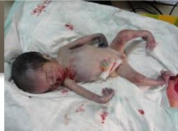
Developmental Periods
- Ovum (zygote): 0 to 14 days
- Embryo: 14 days to 8 weeks
- Fetus: 8 weeks to birth (28 weeks)
- Perinatal period: 28 weeks of gestation to 7 days after birth
Postnatal Periods
- Newborn (neonatal): First 4 weeks after birth
- Infant: Up to one year
- Toddler: 1 to 3 years
Fetal Age Assessment Methods
-
Length measurements
- Crown-heel length
- Crown-rump length
-
Calculation methods
- Up to 5th month: Square root of length of fetus = age in lunar months
- Example: If fetus length is 16 cm, √16 = 4 months
- After 5th month: Length divided by 5 = age in lunar months
- Up to 5th month: Square root of length of fetus = age in lunar months
Head Circumference Development
| Age | Head Circumference |
|---|---|
| Birth | 35 cm |
| 3 months | 40 cm (+5) |
| 1 year | 45 cm (+5) |
| 2 years | 48 cm (+3) |
| 7 years | 50 cm (+2) |
| 10 years | 52 cm (+2) |
Dental Development for Age Determination
Temporary (Primary) Teeth Eruption
| Teeth | Eruption Period |
|---|---|
| Medial incisor (lower) | 6-8 months |
| Medial incisor (upper) | 7-9 months |
| Lateral incisor (lower) | 10-12 months |
| Lateral incisor (upper) | 7-9 months |
| First molar | 12-14 months |
| Canine | 17-18 months |
| Second molar | 20-30 months |
Permanent Teeth
Total: 32 teeth consisting of:
- 8 incisors
- 4 canines
- 8 premolars
- 12 molars
Permanent Teeth Eruption Schedule
| Tooth Type | Eruption Age |
|---|---|
| First molar | 6-7 years |
| Medial incisor | 7-8 years |
| Lateral incisor | 8-9 years |
| First premolar | 9-10 years |
| Second premolar | 10-11 years |
| Canine | 11-12 years |
| Second molar | 12-14 years |
| Third molar (wisdom teeth) | 17-25 years |
Skeletal Development for Age Determination
Bone Ossification Timeline
Early Development (6-15 years)
6 years:
- The pubic ramus of the ischial bone is united with the ischial ramus of the pubic bone
14 years:
- Trochlea is united with the capitulum (humerus)
15 years:
- Both trochlea and capitulum united with shaft of humerus
- Ilium, ischium, and pubis are united to form the acetabulum (disappearance of the Y-shaped suture)
16 years:
- Lateral epicondyle with shaft of humerus
- Upper end of ulna with shaft
- Lesser trochanter of femur with shaft
Later Development (17-25 years)
17 years:
- Medial epicondyle with shaft of humerus
- Upper end of radius with shaft
- Greater trochanter of femur with shaft
18 years:
- Distal ends of metacarpals and proximal ends of proximal phalanges united with their shafts
- Lower ends of tibia and fibula with their shafts
20-21 years:
- Lower ends of radius and ulna with their shafts
21 years:
- Lower end of femur with shaft
- Upper ends of tibia and fibula with shafts
- Ischial tuberosity with ischium
23 years:
- Iliac crest with ileum
- Sternal end of clavicle with shaft
- Basiocciput with basisphenoid at base of skull
Advanced Development (30-70 years)
30 years:
- Closure of sagittal suture (begins from its inner aspect)
40 years:
- Xiphoid process with body of sternum
- Closure of coronal suture
50 years:
- Greater cornu of hyoid bone with its body
- Closure of lambdoid suture
60 years:
- Manubrium sterni with body of sternum
70 years:
- Closure of all skull sutures except temporoparietal
Detailed Ossification CentersY
Humerus:
- Head: Appears at 1 year, Fusion at 14-16 years (female), 14-18 years (male)
- Greater tubercle: Appears at 7 months, Fusion (with head) 2-4 years
- Lesser tubercle: Appears at 7 months, Fusion (with greater tubercle) 5-7 years
- Trochlea: Appears 7-10 years (female), 9-11 years (male), Fusion 9-13 years (female), 11-15 years (male)
Radius:
- Head: Appears at 6 years (female), 8 years (male), Fusion at 14 years (female), 16 years (male)
- Distal end: Appears at 1 year, Fusion at 16.5 years (female), 18 years (male)
Ulna:
- Olecranon: Appears 9-12 years (female), 11-13 years (male), Fusion at 15 years (female), 17 years (male)
- Distal end: Appears 8-10 years (female), 10-11 years (male), Fusion at 17 years (female), 18 years (male)
Femur:
- Head: Appears at 1 year, Fusion at 14-15 years (female), 16-17 years (male)
- Greater trochanter: Appears at 3 years, Fusion at 14 years (female), 17 years (male)
- Lesser trochanter: Appears at 1 year, Fusion at 15-17 years (both sexes)
- Distal end: Appears before birth, Fusion at 14-17 years (both sexes)
Tibia:
- Proximal end: Appears shortly before birth, Fusion at 14-15 years (female), 16-17 years (male)
- Distal end: Appears at 1 year, Fusion at 14.1-14.4 years (female), 16 years (male)
Fibula:
- Proximal end: Appears at 2 years (female), 4 years (male), Fusion at 14-16 years (both sexes)
- Distal end: Appears at 1 year, Fusion at 13-15 years (female), 14-16 years (male)
Medicolegal Age Determination Significance
Legal Age Milestones
6 years
- Age of education begins
- Identification marker: Central incisors eruption
7 years
- Age of discrimination (التمييز):
- Crimes below this age: responsibility of guardian
- Crimes above this age: trial in Child Court but no punishment (until age 12) . Identification: eruption of central incisors
12 years
- Below this age: Child training for work is illegal
- Above this age: Partially responsible (punished but not imprisoned until age 15)
- Identification marker: Eruption of 2nd molar
14 years
- Below this age: Cannot commit the crime of rape
15 years
- End of mother’s custody (الحضانة) for both son and daughter
- Children can choose their custodian until age 21 for son or marriage for daughter
- Above this age: Sentenced as adults with reduced sanctions (no execution, life sentence, hard labor, or aggravated prison)
- Identification markers: Union of trochlea and capitulum with lower end of humerus + acetabular suture union
16 years
- Identity cards issued for both sexes
- Male identification: Union of lateral epicondyle with shaft of humerus
- Female identification: Union of metacarpals with their distal ends
18 years
- Full criminal responsibility begins (ببدا يحكم عليه)
- Age of majority (انكما الابعث الاكما = 21y in some jurisdictions)
Forensic Identification Techniques
Categories of Forensic Science
Biological Forensics
-
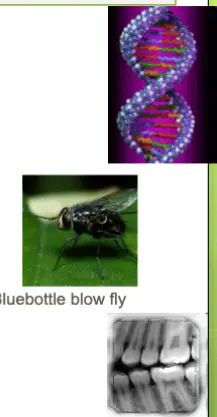
DNA Evidence
-
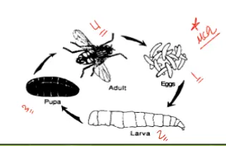
Forensic Entomology
- Study of insects on decomposing remains
- Helps determine time since death (post-mortem interval)

-
Forensic Serology
- Analysis of blood and bodily fluids
- Blood typing and protein markers
-
Forensic Odontology
- Dental identification and bite mark analysis
- Useful for identifying decomposed remains
Forensic Pathology.
• Forensic Psychiatry.
• Forensic Toxicology.
• Bloodstain Pattern Analysis.
• Forensic Photography

Physical Forensics
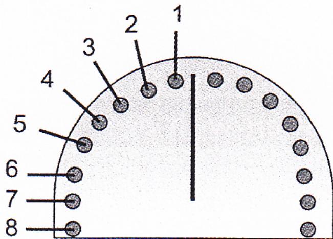
Principles of fingerprint identification (Dactylography) The skin in the palmar aspect of the hand is covered with ridges on which sweat pores open. These ridges are with several types and patterns, which were found to be unique in each individual (l in 64 billion chances for 2 prints to be identical
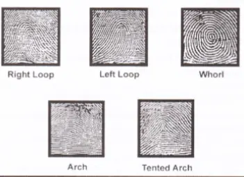
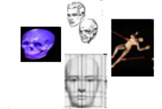
Forensic anthropology

- Forensic Art
- Composite sketching from witness descriptions
- Image modification and enhancement
- Age progression for missing persons
- Post-mortem reconstruction
- Demonstrative evidence for court presentations
Molecular Forensics
Use of identifying characteristics of molecules in our cells to aid legal investigations -
Compare the DNA and/or blood type of crime scene evidence to that of suspects, or use this information to identify a victim.
Fingerprint Identification – The ridges that run through both the dermis and epidermis create a pattern that remains unchanged throughout a person’s life.
- Reversible atrophy of these ridges can occur in certain diseases such as dermatitis.
- Permanent impairment results from conditions like leprosy or after exposure to radiation.
- Attempts to mutilate fingerprints are sometimes made; however, if only the epidermis is destroyed, the ridge DNA remains intact.
DNA Profiling – The following elements are central to its medico‑legal importance:
- DNA
- Gene
- Locus
- Chromosomes
- Mitochondrial DNA