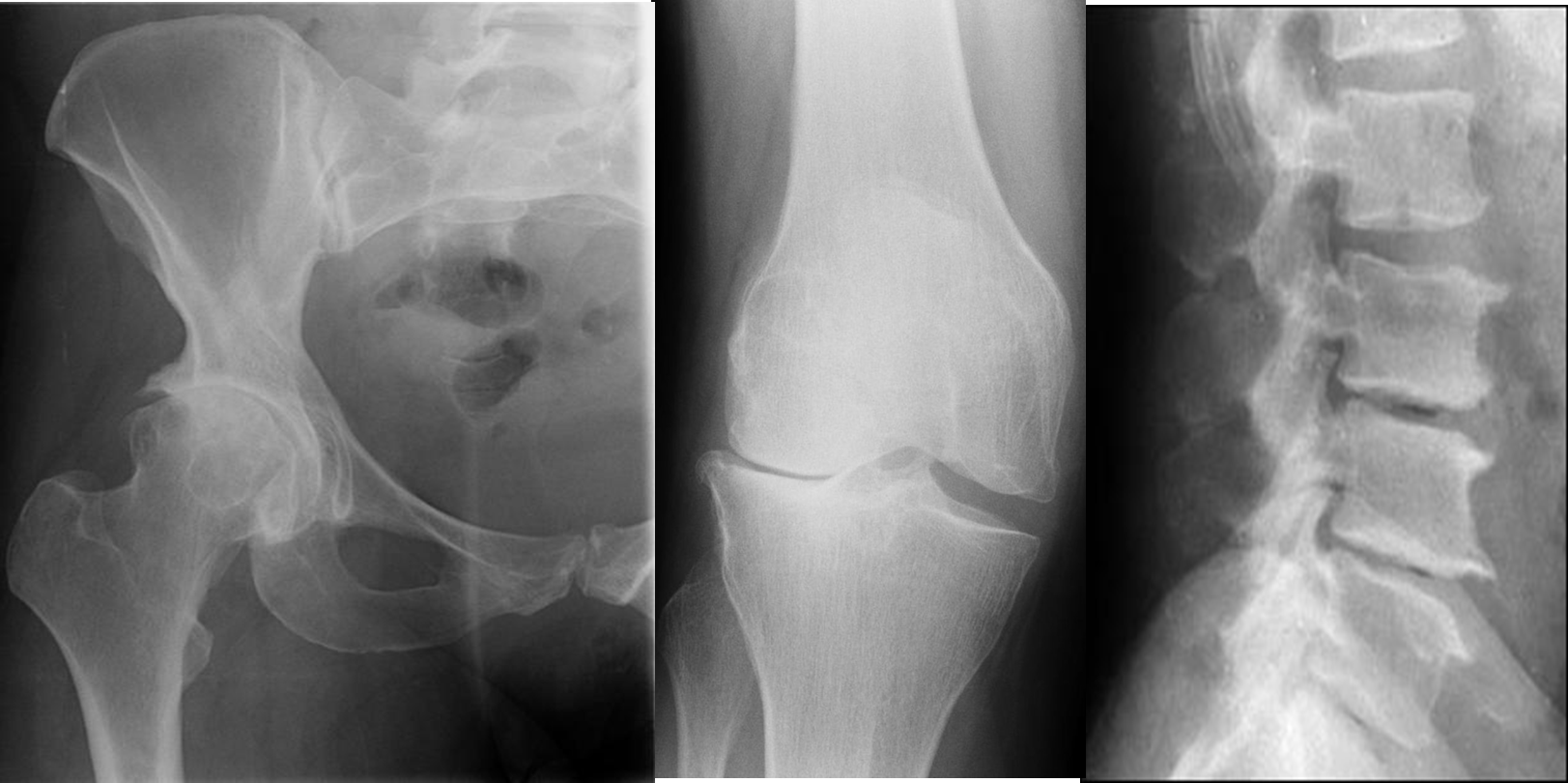
Radiological signs of osteoarthritis
-
Irregular joint space narrowing
-
Subchondral sclerosis: a dense area of bone (visible on x-ray) just below the cartilage zone of a joint.
-
Osteophytes (bone spurs): spurs or densifications that develop on the edges of the joint.
-
Subchondral cyst: a fluid-filled cyst that develops on the surface of a joint

-
Lateral compartment osteoarthritis of the knee X-ray right knee (AP view) Marked lateral compartment narrowing is accompanied by subchondral sclerosis and osteophyte formation. Tibial spine osteophytes are also visible.
-
Degenerative disease of spine with osteophyte formation and vacuum disc phenomenon.